


Clinical Image Article
Nevus of Ota
Department of Dermatology, University Hospital, Hassan II, Fez, Morocco
*Corresponding author: EL Jouari Ouiame, Department of Dermatology, University Hospital, Hassan II, Fez, Morocco, E-mail: eljouariouiame@gmail.com
Received: August 29, 2018 Accepted: September 24, 2018 Published: September 28, 2018
Citation: El Jouari O, Senhaji G, Elloudi S, Baybay H, Mernissi FZ. Nevus of Ota. Madridge J Dermatol Res. 2018; 3(2): 77-78. doi: 10.18689/mjdr-1000118
Copyright: © 2018 The Author(s). This work is licensed under a Creative Commons Attribution 4.0 International License, which permits unrestricted use, distribution, and reproduction in any medium, provided the original work is properly cited.
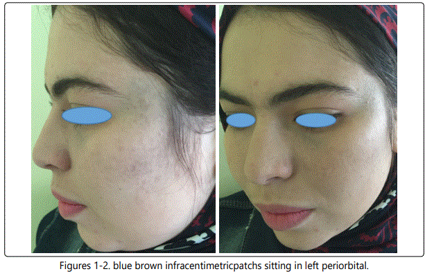
A 32 year old women has had, since birth, pigmented spots of the left periorbital area without involving the sclera [Figure 1, 2].
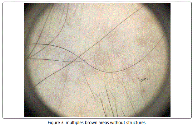
There has been no change in the extent of the lesion. The patient noticed that the spots become sometimes some what darker. Dermoscopy revealed multiples brown areas without structures [Figure 3].
Ota is a congenital pigmentary disorder of the skin and mucus membranes, observed frequently in the yellow race and blacks [1]. It is a macular discoloration of the middle and upper portions of the face with common involvement of the periorbital area [2]. Most cases are sporadic, rare family clusters have been reported [3] and are characterized as being unilateral (95%, having a female predilection (80%, displaying scleral lesions (65%), and being congenital (60%) [2]. The lesions were composed of brown and blue macules the size of wich varied from that of a grain of rice to that of lentil. The overall pigment picture varied from deep purplisch-blue to light brownish-blue [1]. Lesions tend to be slate-gray to brown in color with a ‘powder-blast burn' appearance. The forehead, temple, periorbital area, cheek, and nose are commonly involved. Rarely, pigmentation is bilateral and large areas of the face and oral mucous membranes are affected [3]. The nevus of Ota is a misnomer because it does not contain true nevus cells and derives its pigmentation from dendritic melanocytes which are found in the reticular layer of the dermis [2]. The treatment options for this condition included cryotherapy, dermabrasion, surgical excision and cosmetic camouflage. Although The Q-switched lasers have been used successfully to treat nevus of Ota [4].
Conflict of Interest
The authors declare no conflicts of interest.
References
Contact us for any additional information - contact@madridge.org
 Madridge Publishers is licensed under a Creative Commons Attribution 4.0 International License.