


Research Article
Epidemiology of Dermatophyte Infections in Mongolia. A Five-Year Retrospective Study
1Department of Dermatology, Mongolian National University of Medical Sciences, Ulaanbaatar, Mongolia
2Mycological Laboratory, The National Dermatology Center of Mongolia, Ulaanbaatar, Mongolia
3Department of Microbiology, Mongolian National University of Medical Sciences, Ulaanbaatar, Mongolia
*Corresponding author: Enkhtur Yadamsuren, Department of Dermatology Mongolian National University of Medical Sciences, Ulaanbaatar 14210 Mongolia, E-mail: enkhtur@mnums.edu.mn
Received: November 22, 2021 Accepted: December 02, 2021 Published: December 08, 2021
Citation: Yadamsuren E, Tseden-Ish U, Soyolbaatar B, Jamyansuren E, Byambaa S, Manibadraa O, Chimeddorj B. Epidemiology of Dermatophyte Infections in Mongolia. A Five-Year Retrospective Study. Madridge J Dermatol Res. 2021; 5(1): 106-109. doi: 10.18689/mjdr-1000128
Copyright: © 2021 The Author(s). This work is licensed under a Creative Commons Attribution 4.0 International License, which permits unrestricted use, distribution, and reproduction in any medium, provided the original work is properly cited.
Abstract
Objectives: Dermatophyte infections have been considered to be a major public health problem in many parts of the world. We investigated the epidemiology of fungal skin infections through a retrospective analysis of patientʼs medical records between 2014 and 2019.
Methods: A total of 24,484 patients clinically suspected of cutaneous fungal infections were examined. Skin scales, plucked hairs, nail clippings and sub-ungual debris were examined by direct microscopy and culture. Dermatophyte species were identified atthe species level by a combination of morphological and physiological criteria.
Results: 13,386 (51.0%) of 24,484 participants were male and 12,843 (48.9%) were females. The male to female ratio was 1:1,1. The mean (±SD) and the median age of the participants were 23.5 (±0.12). Direct microscopy confirmed a combinated rate of 28.5% (6,973/24,484 cases). Among the 1747 mycological suspects cases, 560 (32.1%) patients had dermatophyte and yeast infections based on culture. Tinea corporis 2,420 (9.5%) was the most prevalent type of dermatophytosis followed by tinea faciei 1,338 (5.5%), tinea capitis 1,187 (4.9%), and tinea pedis 833 (3.4%). The percentage of positive cultures most frequent isolated was Trichophyton 19 (1.1%), Microsporum canis 7(0.4%), Candida albicans 518 (29.6%), Candida krusei 11(0.6%).
Keywords: Mongolia, Epidemiology, Dermatophyte, Yeast, Skin Infections
Introduction
There is a total of more than 120,000 fungal species in the world, and certainly, a number of them can cause fungal diseases in humans, animals, and plants [1]. Dermatophytosis is an infection of keratinized tissue, including the skin, hair, and nails caused by various types of dermatophyte [2-4]. Dermatophyte infections are very common worldwide [2, 5]. They are believed to affect 20-25% of the worldʼs population [6-10]. Furthermore, the location of infection and type of organism depends on the geographical environment [3, 7, 8, 10-15]. The cause for this increase in the prevalence of skin mycoses possibly may be found in the publicʼs socioeconomic status as well as, poor hygiene and sanitary conditions [16]. The etiologic agents of dermatophytosis (ringworm) are classified into three genera: Epidermophyton, Microsporum, and Trichophyton. Dermatophytosis can occur in any age group and it is found that anthropophilic and zoophilic fungal disease is more common among children and adolescents [15]. The trend of living in communities, contact with animals, the use of -antibiotics, corticosteroids, and antineoplastic drugs are some of the factors that contribute to the increase in the risk of infection by fungi especially by dermatophytes [16]. In 1965, 1987, 2006, Mongolian researchers studied dermatophyte clinical manifestations and epidemiology prevalence, and were zoophilic dermatophyte infection dominant of this time [17-19]. As the epidemiology of dermatophytosis is changing over time it is important to review periodically the incidence of dermatophytes and their distribution [2]. According to many researchersʼ studies, the prevalence of fungal infections has been studied for a 5-10 years period. In our country, it is necessary to study the causative agents of dermatophytes in recent years. For these reasons, the study of the dermatophyte in Ulaanbaatar was initiated.We investigated the epidemiology of fungal skin infections through a retrospective analysis of patientʼs medical records between 2014 and 2019.
Materials and Methods
This study was a retrospective analysis of the data obtained from records of the mycology laboratory at the National Dermatology Center of Mongolia, samples processed from January 2014 through December 2019. A total of 24,484 samples were received during the 5-year study period. Samples of skin, hair, and nails were taken from patients using scalpels, forceps, and glass slides that had been washed in ethanol and sterilized with a Bunsen burner. Direct microscopic observation of the samples was carried out by examining the material in 10% KOH. Clinical specimens were cultured on Petri dishes of Sabouraud dextrose agar (SDA) (Difco, Detroit, MI, USA) containing 50 mgl−1 each of chloramphenicol and cycloheximide (SCC). The inoculated Petri dishes were incubated at 25 °C and examined after 7, 14, 21, and 28 days. Positive cultures were sub-cultured on plates of SCC. The colonies were studied to determine their morphological and microscopic characteristics. Yeast were cultured in Sabouraud agar medium with chloramphenicol for 72 hours. The data were compared using analyses of variance and the Chi-square (χ2) test as appropriate. The level of statistical significance was set at P<0.05. All statistical analyses were performed using Stata /IC 12 program/.
Result
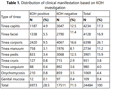
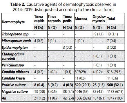
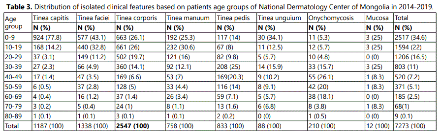
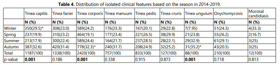
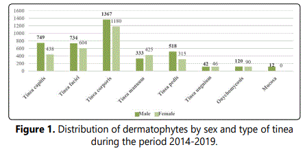
A total of 24,484 samples were received during the 5-year study period. 13,386 (51.0%) of 24,484 participants were male and 12,843 (48.9%) were females. The male to female ratio was 1:1,1. The age range was 1-89 years, andthe mean (±SD) and the median age of the participants were 23.5 (±0.12). The frequency rate of the dermatophyte species in males was higher than in females. Table 1 shows that direct microscopy confirmed a combined rate of 28.5% (6,973/24,484 cases). Tinea corporis 2,420 (9.5%) was the most prevalent type of dermatophytosis followed by tinea faciei 1,338 (5.5%), tinea capitis 1,187 (4.9%), and tinea pedis 833 (3.4%). Figure 1 shows the distribution of dermatophytosis based on the gender of patients, with an evident predominance of women for tinea unguium and manuum and especially tinea corporis more commonly affected males. Among the 1747 mycological suspects cases, 560 (32.1%) patients had dermatophyte and yeast infections based on culture. The percentage of positive cultures most frequent isolated was Candida albicans 518 (29.6%), Candida krusei 11 (0.6%), Trichophytonspp 19 (1.1%), Microsporum canis 7 (0.4%), Epidermophyton 3 (0.2%). Candida albicans 518 (29.6%) was the main etiological agent of the mucosal candidiasis. Trichophyton spp 19 (1.1%) was a dominant agent of the onychomycosis, and Microsporum canis 7 (0.4%) was the etiological agent of the tinea capitis. Epidermophyton 3 (0.2%) was the etiological agent of the tinea pedis. (Table 2) Distribution of isolated clinical features based on patients age group of National Dermatology Center of Mongolia in 2014-2019 shown that table 3. The majority of infections occurred in the age group of 0-9 years 2,517 (34.6%) and 10-19 years 1594 (22%) during 5 years. Tinea pedis was mainly identified in the group of patients aged 30- 39 years 208 (25%). Onychomycosis identifies in the group of patients aged 40-49 years 55 (26.1%). The most identified clinical features were tinea corporis 2547 (35.1%). Table 4 shown that distribution of isolated clinical features based on the season in 2014-2019. Tinea capitis occurred predominantly on winter 350 (29.5%), (P < 0.001). Tinea corporis 778 (32.3%), tinea unguium 31 (35.2%) were occurred on autumn, (P < 0.001).
Discussion
The dermatophyte species of superficial fungal infections differ in different geographical areas and changes over time. In our survey, direct microscopy confirmed a combined rate of 28.5% (6,973/24,484 cases). Tinea corporis 2,420 (9.5%) was the most prevalent type of dermatophytosis followed by tinea faciei 1,338 (5.5%), tinea capitis 1,187 (4.9%), and tinea pedis 833 (3.4%). The majority of infections occurred in the age group of 0-9 years 2,517 (34.6%) and 10-19 years 1594 (22%) during 5 years. Epidemiology of dermatophytosis in Teran, direct microscopy confirmed a contamination rate of 19.7% (2622/13,312 cases) of which 1535 cases (58.5%) were culture positive distributed in male (1022 cases) and female (513 cases), during 2010-2014. The most commonly infected age group was the 30-39 years old. Tinea pedis (30.4%) was the most prevalent type of dermatophytosis followed by tinea cruris (29.8%) and tinea corporis (15.8%) [3]. These differences may be attributed to the geography, regional climate, or population migration, among other factors, in the regions studied. In southwest Poland, from 2468 patients, 2753 fungi were identified including dermatophytes, yeast and moulds, years 2003-2007. Among the yeast-like fungi, a marked predominance of Candida species was observed (86.3%).[10] Similarly, there are reports of frequency of dermatophytosis and yeast. In Swiss, from 1993 to 2000, the total number of samples sent for mycological analysis was 33,725. Dermatophytes were isolated from 4,193 collected samples. Yeast and contaminant moulds were found in 3429 and 2423 cultures, respectively. T.rubrum was the most frequently isolated species accounting for 62.1% of the strains followed by T. mentagrophytes (24.5%) and M. canis (5.0%). Similarly, there are reports in frequency of yeast.[20] In Asia, specially in Guangdong, Southern China tinea was the most common superficial infection. Over the 10-year period, a total of 3385 specimens yielded 697 fungal strains in culture. Tinea unguium was the most prevalent type of superficial infections at 28.55% (199/697, p≥0.05), followed by tinea capitis (15.64 %, 109/697), tinea pedis (15.06 %, 105/697), tinea cruris (10.33 %, 72/697) and tinea corporis (8.75 %, 61/697). T.rubrum was the most common dermatophyte pathogen (56.24 %, 392/697, p≥0.05) both in male and female populations, followed by T. mentagrophytes (13.35 %, 93/697) and M. canis (10.19 %, 71/697).[21] T.rubrum, T.mentagrophytes, and M.canis were the three most commonly isolated species were different from our study.
Conclusion
In our survey, the percentage of positive cultures most frequent isolated was Candida albicans 518 (29.6%), Candida krusei 11 (0.6%), Trichophyton spp 19 (1.1%), Microsporum canis 7 (0.4%), Epidermophyton 3 (0.2%). Our data provide a valuable baseline on which to assess future efforts directed toward the prevention of dermatophytosis infections in our epidemiological setting. Unfortunately, only very sporadically effective measures for the sanitation of known infection sources derived from this literature, and research in this area is still needed.
References
- Eymann C., Wachlin G., Albrecht D., et al. Exoproteome Analysis of Human Pathogenic Dermatophyte Species and Identification of Immunoreactive Proteins. Proteomics Clin Appl. 2018; 12(6): e1800007. doi: 10.1002/prca.201800007
- Seebacher, C., J. P. Bouchara, B. Mignon. Updates on the epidemiology of dermatophyte infections. Mycopathologia. 2008; 166(5-6): 335-352. doi: 10.1007/s11046-008-9100-9
- S Zamani, G Sadeghi, F Yazdinia, et al. Epidemiological trends of dermatophytosis in Tehran, Iran: A five-year retrospective study. Journal de mycologie medicale. 2016; 26(4): 351-358. doi: 10.1016/j.mycmed.2016.06.007
- Degreef H. Clinical forms of dermatophytosis (ringworm infection). Mycopathologia. 2008; 166(5-6): 257-265. doi: 10.1007/s11046-008-9101-8
- Kastelan M., Gudelj U V, Massari L P, et al. Dermatophyte Infections in Primorsko-Goranska County, Croatia: a 21-year Survey. Acta Dermatovenerol Croat. 2014; 22(3): 175-179.
- Sadeghi G., Abouei M., Alirezaee M., et al. A 4-year survey of dermatomycoses in Tehran from 2006 to 2009. Journal de mycologie médicale. 2011; 21(4): 260-265. doi: 10.1016/j.mycmed.2011.10.001
- Ameen M. Epidemiology of superficial fungal infections. Clinics in dermatology. 2010; 28(2): 197-201. doi: 10.1016/j.clindermatol.2009.12.005
- Maraki S, Nioti E, Mantadakis E, et al. A 7-year survey of dermatophytoses in Crete, Greece. Mycoses. 2007; 50(6): 481-484. doi: 10.1111/j.1439-0507.2007.01403.x
- Asticcioli S, Di Silverio A, Sacco L, et al. Dermatophyte infections in patients attending a tertiary care hospital in northern Italy. New Microbiol. 2008; 31(4): 543-548.
- Jankowska‐Konsur A, Dyląg M, Hryncewicz-Gwozdz A et al. A 5-year survey of dermatomycoses in southwest Poland, years 2003–2007. Mycoses. 2011; 54(2): 162-167. doi: 10.1111/j.1439-0507.2009.01774.x
- Panasiti V, Devirgiliis V, Borroni R G, et al. Epidemiology of dermatophytic infections in Rome, Italy: a retrospective study from 2002 to 2004. Medical Mycology. 2007; 45(1): 57-60. doi: 10.1080/13693780601028683
- Koussidou-Eremondi T, Devliotou-Panagiotidou D, Mourellou-Tsatsou O et al. Epidemiology of dermatomycoses in children living in Northern Greece 1996–2000. Mycoses. 2005; 48(1): 11-16. doi: 10.1111/j.1439-0507.2004.01067.x
- Wlodek C, Trickey A, D de Berker et al. Trends in laboratory-diagnosed onychomycosis between 2006 and 2014 in the South West of England. Br J Dermatol. 2017; 176(1): 237-240. doi: 10.1111/bjd.14804
- Reichert-Penetrat S., et al., Epidemiology of dermatophytoses in children living in northeast France: a 5-year study. Pediatric dermatology. 2002; 19(2): 103-105. doi: 10.1046/j.1525-1470.2002.00047.x
- Falahati M., et al., Epidemiology of dermatophytoses in an area south of Tehran, Iran. Mycopathologia. 2003; 156(4): 279-287.
- Monod M, Jaccoud S, Zaugg C, et al. Survey of dermatophyte infections in the Lausanne area Switzerland. Dermatology. 2002; 205(2): 201-203. doi: 10.1159/000063913
- Cai W, Lu C, Li X et al. Epidemiology of superficial fungal infections in Guangdong, southern China: a retrospective study from 2004 to 2014. Mycopathologia. 2016; 181(5-6): 387-395. doi: 10.1007/s11046-016-9986-6
