


Clinical Image
Vitiligo and Alopecia Areata: Association in a Young Man
Department of Dermatology, CHU Hassan II, Fez, Morocco
*Corresponding author: Asmae Rasso, Department of Dermatology, CHU Hassan II, Fez Morocco, E-mail: rassoasmae@gmail.com
Received: July 31, 2019 Accepted: September 24, 2019 Published: October 3, 2019
Citation: Rasso A, Ziani J, Bennnani M, Baybay H, Elloudi S, Mernissi FZ. Vitiligo and Alopecia Areata: Association in a Young Man. Madridge J Dermatol Res. 2019; 4(1): 100-101. doi: 10.18689/mjdr-1000126
Copyright: © 2019 The Author(s). This work is licensed under a Creative Commons Attribution 4.0 International License, which permits unrestricted use, distribution, and reproduction in any medium, provided the original work is properly cited.
Abstract
Vitiligo and alopecia areata (AA) are common autoimmune diseases characterized by white spots on the skin. We report the case of a 63-year-old man, consults for the management of hair removal at the level of beard, Dermatological examination showed vitiligo associated with alopecia areata, this association is rare, whose physiopathology is almost common.
Keywords: Vitiligo; Alopecia areata; Dermoscopy.
Introduction
Vitiligo and alopecia areata (AA) are common autoimmune diseases characterized by white spots on the skin. They have a significant impact on the lives of patients by damaging their appearance and function [1]. The physiopathology is almost common the difference is at the levels of anatomical structures targeted by the immune attack, because the immune attack is directed against the bulb of the dermal hair follicle in alopecia areata and against the melanocytes of the interfollicular epidermis in vitiligo, resulting in a specific distribution of the lesion to the disease. The superficial localization of epidermal melanocytes facilitates their exposure to mechanical lesions that of the “Koebner phenomenon” found in vitiligo, and absent in alopecia areata [1]. These two conditions affect all ethnicities and both sexes, they can occur at any age. In both diseases, the initial source of regeneration is the bulb of the hair follicle (HF), which repopulates the depigmented epidermis of vitiligo with melanocytes [2], and provides melanocytes and keratinocytes for formation, new hair in AA [3].
Image Description
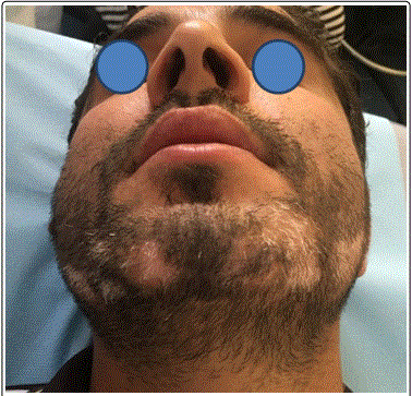
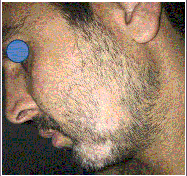
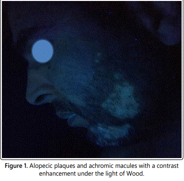
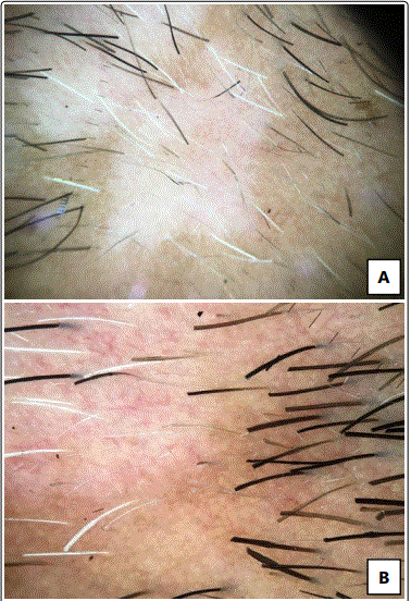
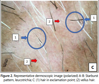
We report the case of 63 years-old man, without significant pathological history, including no autoimmune disease in the family. Consults for the management of depilation at the level of the beard. The dermatology examination revealed non-cicatricial alopecic plaques, and achromic macules at the level of the beard, no squama or erythema, the sign of traction was positive, examination under the light of Wood showed a contrast enhancement (Figure 1), dermoscopy showed a starburst pattern, per follicular depigmentation was predictive of vitiligo, a vellus hair, hair in exclamation point (Figure 2). And then the diagnosis of alopecia areata associated with the vitiligo of the beard was retained. The rest of the clinical examination was normal. The biological assessment in search of other disease autoimmune requested, and did not objectify any other association. The patient was treated with topical tacrolimus with oral corticosteroid low dose with a good clinical improvement after 3 months of treatment.
References
- Barbulescu CC, Goldstein NB, Roop DR, Norris DA, Birlea SA. Harnessing the Power of Regenerative Therapy for Vitiligo and Alopecia Areata. J Invest Dermatol. 2019. doi: 10.1016/j.jid.2019.03.1142
- Goldstein NB, Koster MI, Hoaglin LG, et al. Narrow band ultraviolet B treatment for human vitiligo is associated with proliferation, migration, and differentiation of melanocyte precursors. J Invest Dermatol. 2015; 135(8): 2068-2076. doi: 10.1038/jid.2015.126
- Ito M, Kizawa K, Hamada K, Cotsarelis G. Hair follicle stem cells in the lower bulge form the secondary germ, a biochemically distinct but functionally equivalent progenitor cell population, at the termination of catagen. Differentiation. 2004; 72(9-10): 548-557. doi: 10.1111/j.1432-0436.2004.07209008.x
