Research Article
A Methodological Approach for Analysis of Melanoma Images
Department of Electrical Engineering, Gautam Buddha University, Greater Noida, India
*Corresponding author: Sudhakar Singh, Department of Electrical Engineering, Gautam Buddha University, Greater Noida, India, E-mail: sudhakarsingh86@gmail.com
Received: October 25, 2018 Accepted: November 23, 2018 Published: November 29, 2018
Citation: Singh S, Urooj S. A Methodological Approach for Analysis of Melanoma Images. Madridge J Dermatol Res. 2018; 3(2): 83-87. doi: 10.18689/mjdr-1000121
Copyright: © 2018 The Author(s). This work is licensed under a Creative Commons Attribution 4.0 International License, which permits unrestricted use, distribution, and reproduction in any medium, provided the original work is properly cited.
Abstract
In the proposed work complete task is divided in three parts. First is preprocess and segment the image. After segmentation different features (texture, color, higher order spectra (HOS)) have been extracted. These extracted features are applied for for the classification. Back-propagation-Artificial Neural Network (ANN) has been used for melanoma image classification. Performances of five different algorithms are compared in terms of accuracy, meantime, min-time and max-time. The accuracy of the proposed technique is 84.42% which is beter than other exhisting techniques. In the present work a dataset of 950 images of melanoma is used for the classification in which 500 for training and 250 for testing.
Keywords: Feature Extraction; Back-Propagation-ANN; Melanoma; Skin Cancer.
Introduction
Skin cancer is one of the disease in the modern world which causes death to quite a few number of patients. Medical professionals and researchers are trying to find the root cause of the disease and produce effective medicines for the same. Most of the patients don't know about their skin cancer till it is in final stages. The basic diagnosis of a skin cancer is done using visual inspection by a general physician and if any further investigation needed the case is referred to a dermatologist. A biopsy and pathological analysis is carried out to know the type of cancer (Basal Cell Skin Cancer (BCC), Squamous-Cell Skin Carcinoma and Malignat Melanoma). The deadliest among these three are Malignat Melanoma. It has an assymetrical area, color variation, irregular border and diameter is often greater than 6mm. Maligant Melanoma can cause death if it goes undetected or it spreads in the human skin.
Once the CAD analysis has been done, the skin expert can take the decision based upon the classification results [1]. Dermoscopy is a first and foremost extremely helpful standard technique for diagnosing the spiteful cells of skin disease [2,3]. Skin infection is a large amount added unsafe if it is not found in the premature stages [4].
Related Work
A number of the fuzzy logic techniques examined in preceding research used for color and furthermore skin assessment, including color histogram examination for color marking and skin laceration intolerance [5]. Unfairness, constraint recognition is also defined in skin lesion using fuzzy clustering and texture analysis [5]. The fuzzy clustering segmentation technique is used for the adaptive removing background of skin color for part segmentation [5] and fuzzy c-means clustering considered for pelt scratch analysis [6]. Adaptive fuzzy, c-means improved technique uses local spatial continuity for cluster pattern evaluation [7,8], here skin area segmented by fuzzy estimation grade modeling [8]. Scheduled starting the foundation of artificial intelligence, soft computing, and image processing, used for design as of a number of authors have furthermore, the addition of area understanding concerning the individuality of objects increases the segmentation precision [9,10]. The fuzzy rules are functional among every single point of thoughtfulness (applicant points used for shape detection) single individuals substantial the rule sets are reserved for investigation, by applying a number of criteria of decimation. Based deceitful on the segmented regions of interest, the morphological features and spatial distribution are constructed. These features are classified by a multi-class support vector machine classifier [11,12]. Melanoma parameters including Asymmetry, Border, color, diam, (ABCD) it checked by the lesion Image analysis tools.
Methodology
In the proposed work a set of 950 melanoma images is used. All Images having the same size (1022x762), taken from International Skin Imaging Collaboration [13]. Color features, texture features, HOS features [14] are examined. Back propagation-ANN is used, for performanceanalysis of different algorithms. Block feature, magnitude of patches, angle of patches and normalized feature of cancerous part is calculated. MSE and Accuracy are measured and compared with other existing techniques.
Feature Extraction
Feature selection eliminates the dimensionality of data by selecting only a subset of deliberate features to generate a model. Feature choice is preferable to feature adaptation when the exceptional units and meaning of features are important and the modeling goal is to identify a significant subset.
Color Features
A digit of major color, features has been projected in the literatures, including color histogram [15,16], color moments (CM) [17], color coherence vector (CCV) and color correlogram [18], etc. Among them, CM is one of the simplest yet very effective features. The general moments are mean, standard deviation and skewness; the corresponding calculation can be defined as follows:


Where Xij is the color value of the ith color component of the j-th image pixel and n is the total number of pixels in the image. μi ,σi, γi (i=1,2,3) denote the mean, standard deviation and skewness of each channel of an image respectively [20].
Texture Features
Texture is a quite helpful classification for an extensive series of image. A number of techniques have been projected to remove texture features. Texture features are extracted through computing the pixel statistics or finding the local pixel structures in the original image domain, while the latter transforms an image into the frequency domain and then calculates feature from the transformed image. Gabor filter [19] has been widely used in image texture feature extraction. Thus, texture features can be extracted from this group of energy distributions [20]. Given an input image I (x, y), Gabor wavelet transform convolves I (x, y) with a set of Gabor filters of unlike spatial frequencies as well as orientations. A two-dimensional Gabor function g (x, y) can be defined as follows.

Where σ X and σ y are the scaling parameters of the filter, Wx is the center frequency, and θ determines the orientation of the filter. Figure 1 shows the Gabor function in the spatial domain.
HOS Features
Higher order spectra features are used to investigate the nonlinear and dynamic nature of the given signal [21]. The random distribution of pixels of lesions and nonlinear behavior among the frequency components supports us to use HOS features. Here extracted the HOS features for every 1° of the Radon transform between 0° and 180°. Eq. (5) represents the bispectrum of signal.


Mean of spectral magnitude of power spectrum:

Here X(i) is the Fourier Transform of input signal. Mean of spectral magnitude for HOS.
Contrast

Homogeneity

Back Propagation Neural Network
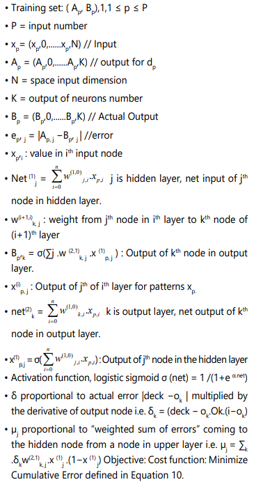
Back propagation is the extended form of the WidrowHoff learning rule in the direction of multiple-layer networks and nonlinear differentiable transfer functions. Input vectors and the analogous object vectors are used to train a network awaiting it can estimate a function, link input vectors by means of specific output vectors. The BPN explained here contains three layers. These layers are input, hidden, and output layers. For the duration of the training phase, the training data are given to the input layer. The data is passed through the hidden layer and then to the output layer. This is called the forward pass of the back propagation algorithm [1].
Algorithm
Back propagation Neural Network algorithm is important for the analysis of image processing. The algorithm is taken from Simon Hykin [22].
Preliminaries


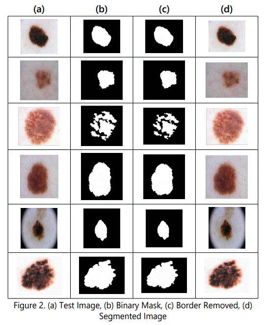

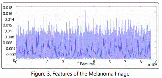
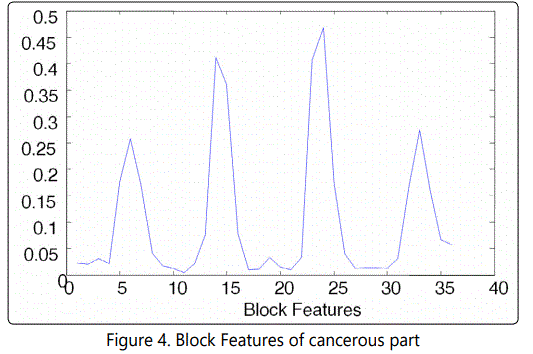
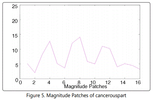
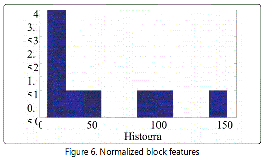
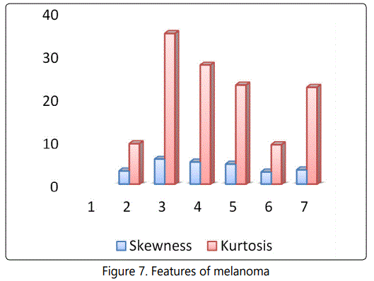
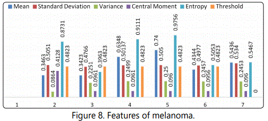

The proposed approach involved in preprocessing, segmentation, feature extraction, feature selection and ANN based classification of the image. The system was tested on a large set of images 950. Primarily, the images used in this study came from different sources. For this reason, care was taken to extract features that are invariant to changes in the imaging conditions. Table 1 shows the statistical features of the different skin diseases. There are six different skin diseases as shown in figure 2 and seven different statistical features are calculated for the classification of skin diseases images. In this paper different feature (color, texture, HOS) for the melanoma image, is discussed. Figure3 depicts texture feature calculated for melanoma image. Figure 4 reprents block features for the melanoma images, these block features are features of cancerous part of the melanoma image. figure 5 represents magnitude Patches of cancerouspart. Figure 6 shows the normalized features of the cancerous part of melanoma images. figure 7,8 brief about statistical features ofmelanoma images. From figure 7 and figure 2 it is observed that the skewness and kurtosis is highest for the second image of column (a), and lowest for first image of column (a). From figure 2 and figure 8, highest value of entropy for image 3 and lowest for the image 2 of column (a). Threshold value of all the images are nearly same. Central Moment of the first image of column is highest. Variance of the third image is highest. Mean valvue of the third image of column is highest.
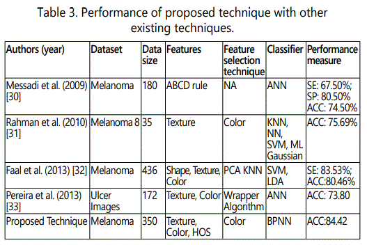
The subsequent table summarizes the results of training this network with the five different algorithms. In all cases, the network is trained up to the error is less than 0.001. The fastest algorithm for this problem is the Polak-Ribiére Conjugate Gradient back propagation algorithm. table 2 shows the performance of the different algorithm used in back propagation ANN. Minimum time taken by the CGP algorithm in terms of meantime, min-time, and max-time. CGP algorithm is better than all four algorithm used. Table 3 shows the performance of the proposed technique with other existing technique. The technique has been used for the 950 melanoma images and accuracy of proposed algorithm is 84.42%, which is better than as compared to the other technique.
Conclusion
In this paper image segmentation, different features (statistical feature, block feature, magnitude features) are extracted and then classification is performed. Performance of different training algorithms are observed in which CGP algorithm was better in terms of time consumption. The accuracy of the thechnique is 84.42% which is better than other three techniques as shown in table 3. In future we plan to incorporate more advanced features related to diagnostic relevance into our system and experiment with other classification techniques.
References