

Clinical Image Article
Dermoscopy of Nevus Spilus
Department of Dermatology, CHU Hassan II, Fez, Morocco
*Corresponding author: Ouiame EL Jouari, Department of Dermatology, CHU Hassan II, Fez, Morocco, E-mail: eljouariouiame@gmail.com
Received: September 5, 2018 Accepted: October 12, 2018 Published: October 19, 2018
Citation: EL Jouari O, Senhaji G, El Mahi H, Gallouj S, Zahra MF. Dermoscopy of Nevus Spilus. Madridge J Dermatol Res. 2018; 3(2): 79-80. doi: 10.18689/mjdr-1000119
Copyright: © 2018 The Author(s). This work is licensed under a Creative Commons Attribution 4.0 International License, which permits unrestricted use, distribution, and reproduction in any medium, provided the original work is properly cited.
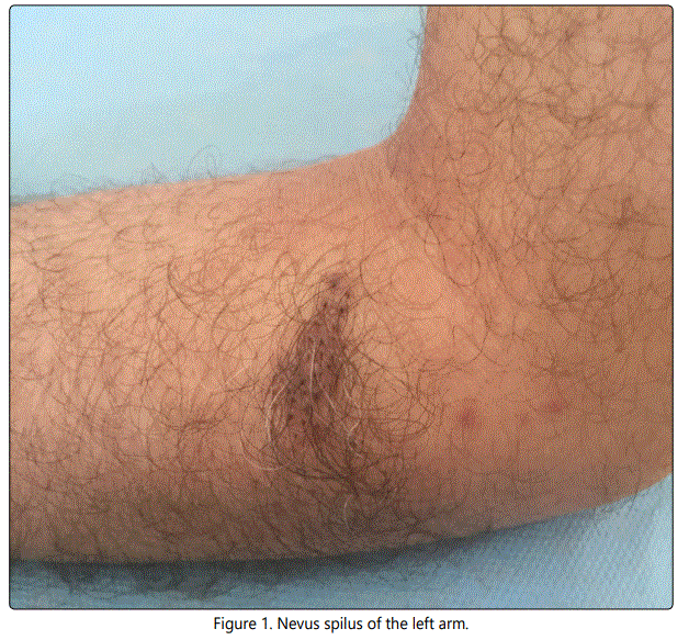
A 60-year-old man had consults for an asymptomatic pigmented macule. The dermatological examination had objectived numerous patchy macules of the left arm, varied in color, ranging from light brownish to brown. Dermoscopy revealed a homogeneous reticular pattern consisted of circular macules and homogenous areas, dots, varying in color from light to dark brown, with hypo pigmented areas.
Nevus spilus also know as speckled lentiginous nevus, spots on a spot, and zosteriform lentiginous nevus [1]. It is a rare dermatologic entity, occurring in 2.8% of examined pigmented lesions. It may be congenital or acquired [2]. Nevus Spilus (NS) is clinically characterized by multiple pigmented macules or papules within a pigmented patch (Figure 1).
It is most frequently located on the trunk, the lower and upper extremities, and the head [3]. Dermoscopy of NS reveals darker brown areas with reticular and globular pattern. The bottom is usually clear and brown lattice. In typical cases of NS, dermoscopy is a reticular pattern with no atypia. In suspected cases of atypical NS, it sometimes reveals a hyper pigmented area with an irregular pattern (Figure 2).

Because of the risk of melanoma by transformation, regular skin examination with the use of dermoscopy is strongly recommended.
Conflict of Interest
The authors declare no conflicts of interest.
References
Contact us for any additional information - contact@madridge.org
 Madridge Publishers is licensed under a Creative Commons Attribution 4.0 International License.