Case Report
Bilateral Pyriform Sinus Fistula: Dermoscopy as an Aid
1Department of Dermatology, Sanjay Gandhi Memorial Hospital, New Delhi-110081, India
2Department of Dermatology & STD, University College of Medical Sciences & GTBH, New Delhi-110095, India
*Corresponding author: Dr. Deepak Jakhar, Department of Dermatology, Sanjay Gandhi Memorial Hospital, New Delhi-110081, India, Tel: +91-9654616205, E-mail: dr.deepakjakhar@yahoo.in
Received: May 14, 2018 Accepted: August 29, 2018 Published: September 3, 2018
Citation: Jakhar D, Kaur I. Bilateral Pyriform Sinus Fistula: Dermoscopy as an Aid. Madridge J Dermatol Res. 2018; 3(2): 75-76. doi: 10.18689/mjdr-1000117
Copyright: © 2018 The Author(s). This work is licensed under a Creative Commons Attribution 4.0 International License, which permits unrestricted use, distribution, and reproduction in any medium, provided the original work is properly cited.
Abstract
Most congenital cervical fistula are derived from first to fourth brachial arches. The skin remnant of fourth brachial arch presents as cutaneous fistula and are termed as pyriform sinus fistula. We present a case of bilateral pyriform sinus fistula in a 7 year old boy and highlight the role of dermoscopy in such cases.
Keywords: Pyriform Sinus Fistula; Fourth Brachial Arch; Cervical Fistula; Dermoscopy Bilateral Pyriform Sinus Fistula; Dermoscopy as an Aid.
Case Report
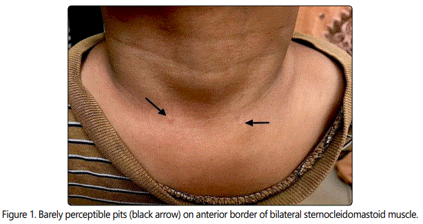
A 7 year old otherwise healthy boy, accompanied by his mother, presented with complaint of on and off serosanguineous discharge from two sides of anterior aspect of neck, just above the sternum, since birth. There was no history of swelling or pain in the past. There was no swallowing or respiratory problems. The child was born at full term by normal vaginal delivery. There was no history of any infections or drug intake by the mother during pregnancy. There was no similar family history. Child had taken oral antibiotics many times in the past but denied any surgical intervention. On examination, two barely visible pits were present on the anterior border of sternocleidomastoid, in close proximity to the sternoclavicular joint [Figure 1].

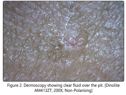
There was no discharge but very subtle erythema at the time of examination. Rest of the muco-cutaneous and systemic examination was normal. Because the pits were barely perceptible, dermoscopy was done in a hope to uncover something which was not visible to naked eyes. Dermoscopy revealed a small pit with very minimal clear fluid [Figure 2].

Given the typical location, clinical features, history since birth and dermoscopic attributes, we kept a provisional diagnosis of bilateral pyriform sinus fistula. Patient was referred to surgery department for further workup and management.
Discussion
Most congenital cervical fistula are derived from first to fourth brachial arches [1]. Near about 95% of cervical anomalies arise from 2nd brachial arch, with fourth brachial arch contributing the least [1]. The skin remnant of fourth brachial arch presenting as cutaneous fistula has been termed as pyriform sinus fistula [1]. An extensive search of english literature revealed only a handful of cases [1-5]. Ohno et al described 7 cases similar to our case but all were left sided [1]. Bilateral involvement has never been reported in english literature to the best of our knowledge. Similar cases with unilateral involvement have been described 4 times in dermatology literature but have been variably referred to as dermoid cyst [2-5]. Ohno et al argued that the possible misdiagnosis in all 4 cases may be because of unfamiliarity of dermatologists to fourth brachial arch fistula in this region. They further argued that a dermoid cyst should have been in the midline rather than on lateral sides [1]. Various authors have stated that the fourth brachial cleft remnant fistula opens on the anterior border of the sternocleidomastoid muscle, possibly at sternoclavicular joint [6, 7]. Liston was first to state the possible existence of this entity and described its theoretical course from apex of pyriform sinus to a skin opening [6].
Bilateral involvement and role of dermoscopy in aiding the diagnosis makes this case unique. We intend to familiarise the dermatologists with this rare entity in a hope that reporting of similar cases in future will help us understand this entity more clearly.
Conflict of Interest
The authors declare no conflicts of interest
References