Clinical Image Article
Dermoscopy of a Corneiform Pyogenic Granuloma
Department of Dermatology, CHU Hassan II, Fez, Morocco
*Corresponding author: Cheymae Saadani Hassani, Department of Dermatology, CHU Hassan II, Fez, Morocco, E-mail: ch.saadani@gmail.com
Received: May 29, 2018 Accepted: June 28, 2018 Published: July 4, 2018
Citation: Hassani CS, Elloudi S, Baybay H, Mernissi FZ. Dermoscopy of a Corneiform Pyogenic Granuloma. Madridge J Dermatol Res. 2018; 3(1): 50-51. doi: 10.18689/mjdr-1000111
Copyright: © 2018 The Author(s). This work is licensed under a Creative Commons Attribution 4.0 International License, which permits unrestricted use, distribution, and reproduction in any medium, provided the original work is properly cited.
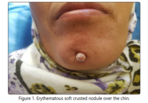
A 30-year-old female presented with 4 months history after a trauma, of an erythematous, soft crusted and painful nodule over the chin (Figure 1).

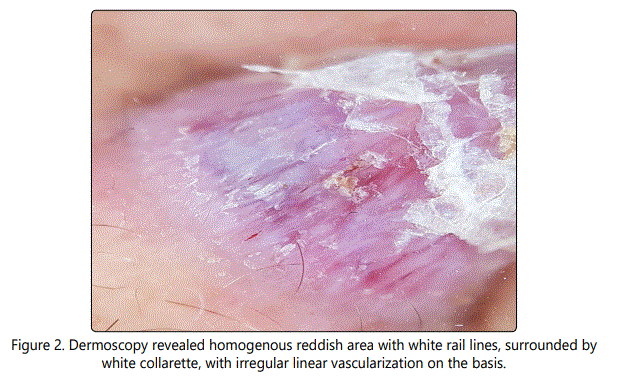
Dermoscopy was performed and revealed homogenous reddish area with white rail lines, an irregular linear vascularization at the basis, and white crusts with collarette at the upper (Figure 2).

Suggestive of pyogenic granuloma. Histopathology confirms the diagnosis revealing lobular capillary proliferation in upper dermis.
Pyogenic granuloma is dermoscopically characterized by reddish homogeneous area which is corresponded to the histologic finding of proliferating capillaries and veins [1]. The white lines like a double rail correspond to the histologic finding of fibrous septa that surround the lobules [2]. And finally, a white collarette corresponds to the hyperplastic adnexal epithelium that embraces the lesion at the periphery [1].
The identification of vascular structures in pyogenic granuloma is crucial because the presence of vascular structures in a red tumor resembling a pyogentic granuloma indicates that a melanoma should not be ruled out [3], such as in our case.
Conflict of Interest
The authors have no conflicts of interest to disclose.
References