Research Article
Ultrasound Analytic Criteria for diagnosing Cutaneous Malignant Melanoma
1Clinic for Dermatology Hautaerzte am Marktplatz, Karlsruhe, Germany / Endovision Medical Center, Yerevan, Armenia
2National Center of Oncology V.A.Fanarjyan, Yerevan, Armenia
*Corresponding author: Michael Hambardzumyan, Clinic for Dermatology, Hautaerzte am Markplatz, Kaiserstraße 72, 76133 Karlsruhe, Germany, E-mail: hambardzumyan.michael@gmail.com
Received: October 8, 2017 Accepted: January 25, 2018 Published: January 31, 2018
Citation: Hambardzumyan M, Hayrapetyan A. Ultrasound Analytic Criteria for diagnosing Cutaneous Malignant Melanoma. Madridge J Dermatol Res. 2018; 3(1): 32-36. doi: 10.18689/mjdr-1000108
Copyright: © 2018 The Author(s). This work is licensed under a Creative Commons Attribution 4.0 International License, which permits unrestricted use, distribution, and reproduction in any medium, provided the original work is properly cited.
Abstract
Multimodal ultrasound analytic criteria can optimize primary cutaneous melanoma features; reveal satellite pathologies or regional lymph node metastases. The most significant malignant melanoma ultrasound criteria are its shape, hypoechoic homogenous structure, retro mass acoustic enhancement, hypervascularization, multiple peduncles, high strain elastography stiffness and high Length/Thickness (L/T) ratio. Ultrasound assessment of the cutaneous melanoma into 3 groups, according to their shape: oval, jagged and superficial types can be useful in the clinical practice. Consideration of the several ultrasound criteria can help with the treatment options for melanoma.
Keywords: Malignant Melanoma; Ultrasound; Analytic Criteria.
Abbreviations: CD: Color Doppler; PD: Power Doppler; ADF: Advances Doppler Flow; L/T: Length/Thickness Ratio; FNA: Fine Needle Aspirations.
Introduction
Cutaneous ultrasound is a tool of great interest in the preoperative study of cutaneous malignant melanoma [1-3]. It identifies thin melanomas fairly accurately and can be useful for planning appropriate surgical margins. Authors [4,5] proposed an automatic diagnostic system based on quantitative analysis of ultrasound data for differential diagnosis of melanocytic skin tumors tested on 160 ultrasound data sets (80 of malignant melanoma and 80 of benign melanocytic nevi). Using parameters, they were able to differentiate malignant melanoma from benign melanocytic skin tumors with 82.4% accuracy (sensitivity = 85.8%, specificity = 79.6%) and to provide supplementary information on lesion penetration depth.
Preoperative high-resolution ultrasound is a noninvasive examination that can help in choosing appropriate surgical margins and should reduce the need for partial or excision biopsy before surgery, and the need for further re-excision [6]. This article illustrates the various aspects of the locoregional spread of cutaneous melanoma, as imaged with grayscale ultrasound and doppler techniques. High-resolution ultrasound allows recognition of small, clinically occult melanomatous foci within the skin and lymph nodes. We discuss the possibilities and limitations of ultrasound in the initial staging (primary melanoma, satellite metastasis, in-transit metastasis, and lymphadenopathy), selection for sentinel lymph node biopsy procedure, patient followup, detection of recurrence, and ultrasound-guided intervention [7].
Ultrasound examination with a 12-15 MHz linear transducer can reliably differentiate the primary melanoma > 1 mm from those < or 1 mm. The sensitivity, specificity, positive and negative predictive values of ultrasound to detect melanoma > 1 mm were 92%, 92%, 95% and 81% respectively. The correlation between ultrasound and histological tumor thickness was very good [Pearson's correlating index 0.823 (P < 0.001)] [8].
Mostly in all hospitals and clinics, the linear highresolution probes from 8MHz to 15 MHz were available. The advantages of high-resolution probes from 15 MHz to 20 MHz or 30 MHz or higher for melanoma tumor thicknesses were discussed in some publications. The aim of this study was to explore cutaneous ultrasound with widely used ranges of linear probes as a tool for evaluation the ultrasound analytic criteria of the malignant melanoma, revealing satellites or distance intradermal pathologies for the optimizing the melanoma patients treatment.
Methods
A total of 30 patients (12 men and 18 women) with a mean age of 62,3 years clinically, dermoscopic and pathologic identified, were included in the study: 24 patients with primary cutaneous melanoma, 1 patient with 2 melanomas with different localizations, 1 patient with primary cutaneous melanoma and satellite dermal mass - totally 26 patients with 27 cutaneous malignant melanomas and 4 patients with metastatic lymph nodes of melanoma. Cutaneous melanoma located on the head and neck 22,2%, on the upper extremities 7,4%, on the trunk 40,8%, on the lower extremities 29,6%. Histological examination of the removed tissues were done in all cases and included the tumor surface, examining its thickness, lymphoid infiltration, mitotic index, nevus association, examinations of the dermal masses surrounded tissues to reaveal the intradermal satellite masses or lymph nodes metastases.
The ultrasound structure of cutaneous tumors and the surrounding tissue was evaluated by Toshiba Xario TUS-X200 system (Toshiba Medical System, Tokyo, Japan) before dermoscopy with DermLite DL3N Dermatoscope (3Gen Inc., San Juan Capistrano, CA, USA) and any clinical procedure like FNA or biopsy. We used 8 MHz probes with extended field of view, zooming and panoramic reconstruction software, improving information quality. In each case, we explored several ultrasound criteria including tumor shape, thickness, length and thickness ratio, acoustic phenomena, echogenesis, echostructure, CD, PD, ADF, tumor peduncles, and Elastography stiffness. To find in melanoma cases the nearby dermal and intradermal satellite pathologies we explored also surrounding soft tissues up to 5cm-7cm from the border and also proximal regional lymph nodes areas. During the cutaneous examination, we rotated the ultrasound probe around its axis in a 180-degree clockwise and counterclockwise to check the entire surface and reveal the maximal diameter and thickness of the melanoma. The ultrasound finding of the deepest part of the melanoma and the visualization of the skin-affected edges can assist dermatologists and pathologists in the histological examination and optimize the surgery. We always took into account the results of clinical examination and dermoscopy.
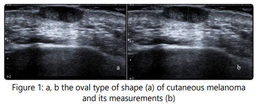
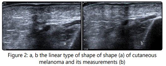
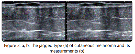
Analyzing all 27 cutaneous ultrasound images we clearly found two main types of melanoma mass shapes: nodular (Fig 1a) and superficial (Fig 2a). Definition and thickness measurements in both groups are quite easy (Fig 1b, 2b). In some superficial or linear shape types of malignant melanoma, we found also intermediate cases with the part of the tumor starting its asymmetric vertical jagged growing (Fig 3a). This jagged type can be in any place of the superficial melanoma: in the middle, in the end, or in one or two parts of the linear shape of the mass. The measurements had to be appropriate and included its deepest part (Fig 3b). Taking into consideration not only the radiological aspects but the clinical point as well, it is more convenient to analyze the cutaneous melanoma group according to 3 shape types of tumor: oval shape, jagged shape, and linear shape. We also registered the ratio between the diameter of the mass and its maximal thickness - L/T ratio.
Results
We divided the ultrasound criteria for 27 malignant melanoma cases into 3 groups (Table 1).
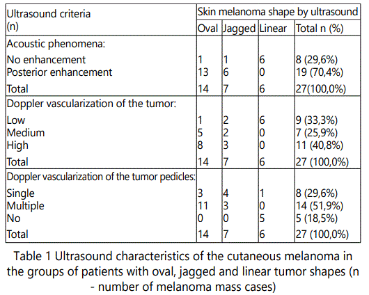
14 patients (51,9%) with an oval shape, 7 patients (25,9%) with jagged shape and 6 patients (22,2%) with linear or superficial tumor shape.
Analyzing ultrasound acoustic phenomena in the cutaneous melanoma group a posterior enhancement in main cases were found (Table 1). The posterior enhancement was quite regular in the oval shape group, less frequent in the jagged and there were no registries in the linear group. Doppler vascularization of the tumor was high in the group of an oval shape, medium in the jagged group and low in linear types of melanoma mass shape. The number of the tumor peduncles vascularization decreased in the group with oval, jagged and linear types of melanoma mass shape.
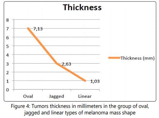
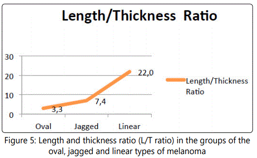
One of the most important characteristics for malignant melanoma is the tumor thickness correlated with tumor shape (Fig 4): it was maximal in the oval group, intermediate in the jagged and low in the linear group. The inverse pattern was noted in L/T ratio: the number was low in the oval group, intermediate in the jagged and maximal in the linear group (Fig 5).
The elastography stiffness ratio has the same tendency: maximal in the oval shape (3,26), interrelate in the jagged (2,76) and minimal (2,40) in the linear shape. The elastography stiffness of cutaneous melanoma and melanoma lymph node metastases of 31 patients was 2,97±0,19, the elastography stiffness of the 27 cutaneous melanoma group was 2,95±0,18 and in 4 cases of the lymph node melanoma metastases group, it was 3,09±0,20. For melanoma lymph node metastases were common oval shape, isoechoic nonhomogeneous structure changing, hypervascularization in CD and PD and ADF.
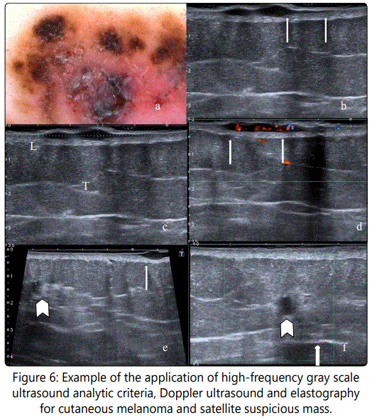
a. A 63 years old woman admitted for ultrasound examination after dermoscopy.
b. Ultrasound analysis: the margin shape was sharp jagged well-defined margin. In the right part of the same mass, it is possible to note the thinnest part of the melanoma tail (arrow) having15, 3mm length and till 0,6mm thickness.
c. The maximal diameter of the mass was 34,5mm and maximal thickness of the mass was 3,2mm. The L/T ratio reached 10, 8 and was higher the ratio for benign pathologies and an average for melanoma (7,9).
d. CD analyses find hypervascularization with disorganized central vessel distribution and also two peduncles. Strain elastography ratio ranges were between 4,20 and 4,94 and it was higher the average for melanoma.
e. During the ultrasound examination of the primary tumor surround tissues, another mass (arrowhead) was founded 42mm left and lateral from the primary skin mass (arrow) and 11-12mm deeper from skin surface. It has an oval shape, 4,2mm diameter, and hypoechoic echogenicity. The L/T relation was 1,1 and the elastography stiffness was high 2, 3.
f. The mentioned criteria are common for the satellite metastases but CD, PD and ADF did not find in the mass the blood flow (arrowhead), some colored dots are out of the mass (arrow). Surgery included removal of both masses. Pathological examination of the skin mass defined superficial Ultrasound of cutaneous melanoma patients also can suspect intra dermal metastases in the surrounded tissue (Fig 6). melanoma with the same thickness noted by ultrasound; the histology of the soft tissues mass was oleogranuloma.
Some ultrasound criteria in gray scale, strain elastography stiffness suspect the possibility of the melanoma satellite metastases; the other ultrasound criteria including CD, PD, and ADF mistrust the metastatic natures of the revealed mass near the primary melanoma. Surgery and pathology confirmed the diagnosis of superficial melanoma in the primary tumor and oleogranuloma in the suspected satellite mass. So, there is no absolute universal criterion for the diagnosis of malignant pathology. It is necessary to use a multimodal approach. In the other rare case, high-frequency ultrasound revealed 2 localisations of skin melanoma in the different parts of the head (Fig. 7).
For outlining the melanoma profile on the skin after the ultrasound we marked the borders with ink to help dermatologist easily configure the optimal skin flap for its removal, as well pathologists, to have the possibility to define the right slices direction for detecting the thickest part of melanoma. Such an approach to the issue can minimize the unnecessary preparation of slices for histological examination.
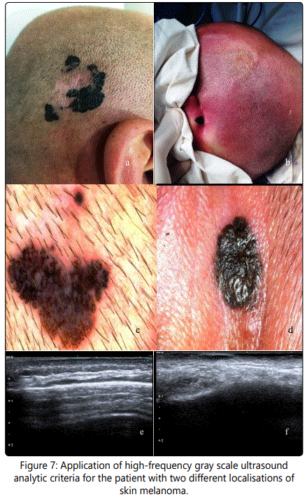
Patient: 38 years old male, with a family history of melanoma. He noted dermal changes near right ear about 5-6 yaers ago (a). Later, (about 1 year ago) the second lesion near the left ear was noticed (b). Before the hospital admission, he had the dermoscopy imaging of the same pathologies near the right ear (c) and left ear (d). Ultrasound examination revealed the linear type of melanoma on the left side (e) with the tumor thickness 0,5mm, L/T ratio 47,6 and on the right side (f) oval shape type with the tumor thickness 1,3mm L/T ratio 6,2. Surgery and pathology were done at the same time for both of the formations and revealed a superficial and nodular type of pigmented melanoma without ulceration, with lymphoid infiltration and high mitosis.
Discussion
For the ultrasound analysis of cutaneous melanoma, we used standard types of linear high-resolution probes, which are available in almost all hospitals and clinics. Ultrasound assessment of malignant melanoma into 3 groups according to their shape into oval, jagged and superficial types can be useful in the clinical practice. Such an approach indirectly confirmed another research: The ultrasound examination can reliably differentiate primary melanoma > 1 mm from those < or 1 mm [8]. In our study, the mean diameter of the linear or superficial shape of melanoma was 1,03mm (Fig. 4).
There are reports in the literature [9] of subclinical satellite lesions that were diagnosed using ultrasound. From the other hand in their pilot study, the other researchers noted the Elastography can enhance the diagnostic accuracy of ultrasound for differentiating between reactive and malignant cutaneous melanoma lymph nodes and might eliminate the need for sentinel lymph node biopsy [10]. Ultrasound provides a detailed preoperative study, reducing the number of incompletely excised lesions and avoiding large resection, which could lead to functional and aesthetic problems [11]. It is an opportune method for revealing the depth invasion of melanoma, farther, it helps in the preoperative staging of melanoma. The measurement of the tumor thickness should be based on the site of greater invasion. It can be useful in the planning surgery. Pre-operative skin marking can help pathologist to specify the melanoma's deepest invasion and its borders in the removed flap tissue, as well it can help in pre-surgical monitoring in pathological changes of the skin.
The most significant melanoma ultrasound criteria are: sharp well-defined margin, hypoechoic mainly homogenous structure, retro mass acoustic enhancement, hypervascularization in different types of Doppler examinations, more than one peduncles, also high L/T relations and high strain elastography stiffness. The results can be discussed according to the resolution of the used linear probes, their pros, and cons, estimation of changes of the superficial type of melanoma to the more invasive or aggressive types of melanoma: jagged and oval shapes.
Conclusion
Ultrasound examination suggests features in malignant melanoma, reveals satellites or intradermal pathologies, lymph node metastases, optimize surgical approaches and can help pathologist with histological examination. In each melanoma case, it is useful to define several ultrasound criteria including tumor shape, thickness, length, thickness ratio, acoustic phenomena, echogenesis, echostructure, color Doppler, power Doppler, advances Doppler Flow of the tumor, its peduncles, and Elastography stiffness. Assessment of all ultrasound criteria by 3 main types into oval, jagged and superficial can more accurately reflect on melanoma skin cancer. Consideration of the different ultrasound criteria helps to establish a melanoma treatment planning.
Conflicts of Interest
The authors have no conflicts of interest to declare.
References