

Review Article
The Role of Magnetic Resonance Imaging in the Diagnosis of Submitral Aneurysm: A Systematic Review
Running Title: Magnetic Resonance Imaging in Submitral Aneurysm
1Cardio-Thoracic Center, Clínica Girassol, Luanda, Angola
2Cardiology Department, Hospital Militar Principal/Instituto Superior, Luanda, Angola
*Corresponding author: Humberto Morais, Military Hospital Main / Higher Institute Rua Pedro Miranda 40-43 Maianga, Luanda Republic of Angola, E-mail: hmorais1@gmail.com
Received: June 1, 2017 Accepted: June 12, 2017 Published: June 19, 2017
Citation: Manuel V, Morais H. The Role of Magnetic Resonance Imaging in the diagnosis of Submitral Aneurysm: A Systematic Review. Madridge J Cardiol. 2017: 2(1): 21-24. doi: 10.18689/mjc-1000106
Copyright: © 2017 The Author(s). This work is licensed under a Creative Commons Attribution 4.0 International License, which permits unrestricted use, distribution, and reproduction in any medium, provided the original work is properly cited.
Abstract
Background: Multimodality imaging has been used in diagnosis of submitral aneurysm, including the use of magnetic resonance imaging.
Objective: This study sought to investigate whether magnetic resonance imaging can be useful in the diagnosis of submitral aneurysm.
Methods: A systematic literature review in PubMed, LILACS and Google Scholar limited to English language with no time restriction was performed. Articles that fulfilled predefined inclusion criteria were included and systematically reviewed according to the PRISMA. Two reviewers independently assessed all titles, abstracts or full text for inclusion.
Results: A total of seven case reports were included. The mean age was 39±20.2 years old. Five were male. Four patients were in Class NYHA II. One had ventricular tachycardia and another one had atrial fibrillation. The diagnosis of submitral aneurysm was done by transthoracic echocardiography in all patients except one. In this case the first hypothesis was mitral endocarditis with an abscess, the magnetic resonance imaging showed a submitral aneurysm. In all cases magnetic resonance imaging confirmed the diagnosis of submitral aneurysm, and in three cases provided additional information.
Conclusions: Magnetic resonance imaging is an excellent non-invasive imaging method in diagnosis of submitral aneurysm when compared to transthoracic echocardiography, and should be indicated in case of doubt in diagnosis, and is useful to answer specific clinical questions.
Keywords: Submitral aneurysm, Magnetic Resonance, Echocardiography, Congenital heart disease.
Abbreviations: LV: Left Ventricle; M: Male; MI: Mitral Insufficiency; MR: Mitral Regurgitation; MRI: Magnetic Resonance Imaging; MV: Mitral Valve; SMA: Submitral Aneurysm; CT: Computed Tomography; TTE: Transthoracic Echocardiography.
Background
Submitral aneurysm (SMA) is a rare and poorly understood cardiac disease that occurs commonly in young African adults, although some cases have been reported worldwide [1-4]. It was also described in pediatric population [5-11].
Submitral aneurysm is considered a congenital disease caused by developmental defect due to weakness at the atrioventricular junction near the posterior mitral annulus. Others etiologies have been reported as tuberculosis, human immunodeficiency virus (HIV), rheumatic carditis, trauma, Takayasu arteritis, cardiac lymphocytosis, Chlamydia pneumoniae in peripartum and also post-myocardial infarction [7,12-15].
According to the extent of posterior mitral annulus involvement by the neck of the aneurysm the SMA is classified in type I - has a single neck, type II – has multiple necks and type III - when the entire posterior mitral annulus is involved [12].
Patients with SMA may be asymptomatic; however, symptoms such as heart failure, arrhythmia, thromboembolism, aneurysm rupture and even sudden death occur do in majority of the patients [12,16,17].
Transthoracic echocardiography (TTE) remains the mainstay for diagnosis of SMA but may have some limitation [18-21]. Over the past two decades, other imaging modalities such as three-dimensional echocardiography, computed tomography (CT), as well as the magnetic resonance imaging (MRI) have been used in diagnosis of SMA [18-28].
MRI has been increasingly used in the evaluation of cardiovascular diseases. A consensus panel on indications of cardiac MRI [29] and guidelines and protocols for cardiac magnetic resonance in children and adults with congenital heart disease were published [30].
The question addressed herein was “What is the role of MRI in the diagnosis of SMA?”
To answer this question we performed this study to investigate whether MRI can be useful in the diagnosis of SMA and if MRI provides additional information.
Methods
We searched the PubMed, LILACS and Google Scholar databases through August 20, 2016 using the MeSH term submitral aneurysm according to the Preferred Reporting Items for Systematic Reviews and Meta-Analyses statement, PRISMA [31] Two investigators working independently screened all titles and abstracts of the studies for eligibility. Duplicate citations were discarded after the preliminary search results were obtained. Furthermore, the references lists of all included studies and relevant review studies were also manually searched.
Inclusion Criteria
Case reports, original research studies, randomized control trials or meta-analyses in humans having data regarding patients with submitral aneurysm diagnosed by TTE and MRI were included.
Exclusion criteria
The exclusion criteria were as follows: Non-English language articles, review articles, papers describing study designs, articles that reports patients with SMA diagnosed by TTE but not with MRI or do not report patients with SMA. Articles with no available full-text as well as editorials or letters to the editor without original data were also excluded.
Data extraction
The following information and data were extracted from included studies: name of the first author, year of publication, country of origin, number of participants, participantsʼ ages and sexes, findings of TTE and findings of MRI were examined and critically analyzed.
Quality Appraisal
The quality appraisal was established according to Le Floch and colleagues criteria. This tool appraises the quality of the study based on the following questions: Did this article give an answer to the research question? Did the article focus clearly on the research question? Was the methodology appropriate? Do you believe the results? (Can it be due to chance, bias or confounding?). To be included, the article had to score “yes” on every question [32].
Results
Study selection
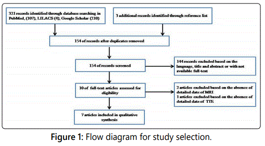
A flow diagram of study selection is shown in Figure 1. A total of 324 papers were initially identified, of which 314 were excluded for not meeting the inclusion criteria. After review of the full texts of the 10 remaining studies, two were excluded based on the absence of detailed date of MRI, and one based on the absence of TTE date. Thus, seven articles published between 2005 and 2016 were included in this systematic review [22-28].
Study description and patients characteristics
Study characteristics and patientʼs characteristics are summarized in Tables 1. A total of seven case reports were included in this study. Two cases were from India and two from Belgium, one from Taiwan, Switzerland, and the last one from Angola. The mean age was 39±20.2 years old, five were male and two were female. Four patients were in class NYHA II, two in NYHA III and one was in NYHA I. Two of them had arrhythmia, one had ventricular tachycardia and one had atrial fibrillation. All patients underwent TTE and MRI.
TTE and MRI findings
The TTE and MR findings are summarized in tables 1. The diagnosis of SMA by MRI was done correctly in all cases, while the diagnostic of SMA was made by TTE in six out of seven patients. In the case of the misdiagnosis, the first diagnostic hypothesis based on TTE was mitral endocarditis with abscess, the MRI showed a SMA [24].
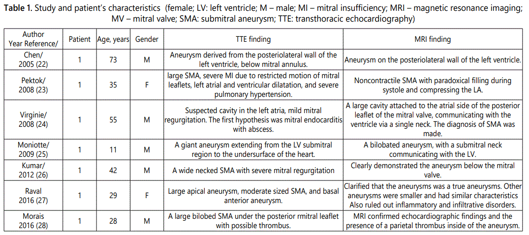
Regarding specific questions - in one case the doubt was the presence of thrombus inside of the aneurysm, the presence of thrombus was confirmed by the MRI [28]. In other case the doubt was if the aneurysm was a true aneurysm or a pseudoaneurysm, the MRI showed that was a true aneurysm [27]. Finally, in one patient the TTE showed a huge SMA and the MRI showed a bilobed SMA, with a submitral neck communicating with the LV, thus improving the anatomic delineation of the aneurysm [28].
Discussion
The questions that should be answered with the available imaging modalities for diagnosis of SMA concern the exact localization, and size of aneurysm, size of basic wall defect, spatial relationship and potential involvement of the mitral apparatus, morphological features of the aneurysm in the proper sense of true or pseudoaneurysm, assessment of global LV function, mitral regurgitation (MR) and potential adverse hemodynamic effects of the aneurysm [18-28].
The TTE remains the first-line investigation for diagnosis of SMA; however, it may have some limitation to respond all these questions [18-21,24,28,33]. In the present review, the diagnosis of SMA was correctly done by TTE in 6 of 7 patients, when compared with MRI which was correct in 7 out of 7 patients.
Misdiagnosis or doubt in diagnosis of SMA by TTE is not rare, even in experienced center. Manuel et al reported a case of a child in which the preoperative diagnosis was mitral valve aneurysm with severe MR but the diagnosis of SMA was made only intraoperatively [7]. Essop et al reported that TTE was unable to show the origin and location of the neck of the aneurysm in two out of five patients with SMA [33]. Moreover, in the present review we present a case reported by Virginie et al, in-which the initial diagnosis was mitral endocarditis with abscess, and the final diagnosis of SMA was clarified by MRI [24].
Recently, MRI has emerged as the preferred non-invasive technique for assessment of LV shape, the degree of aneurysm thinning, and respectability [34,35]. MRI can distinguish between pericardium, thrombus, and myocardium, and helps resolve uncertainty between a true and a pseudoaneurysm [36,37]. In the case of a true aneurysm, the tissue making up the wall of the aneurysm shows delayed myocardial enhancement. As the wall of a pseudoaneurysm is composed only of pericardium, it shows no delayed myocardial enhancement; however, its border does show enhancement. Pseudoaneurysm may show delayed pericardial enhancement.
In present review the MRI showed an intracavitary thrombus that was missed in TTE. It also allowed accurate identification of true aneurysm and provided the necessary information, regarding MV anatomy, grade and mechanism of MR.
Taking into account the aforementioned limitations of TTE, several studies showed the superiority of transesophageal echocardiography, real-time tridimensional echocardiography and CT when compared with TTE [18,21,33].
In contrast, there is no study comparing the performance of MRI with TTE in diagnosis of SMA. The MRI has some limitations such as: availability scan time duration, expense and lack of portability when compared to TTE [38].
Our systematic review can contribute to clarify the value of MRI in the particular diagnosis of SMA. We believe that MRI should be applied as a second diagnostic method in order to corroborate or to dismiss the echocardiographic findings before making clinical decisions in SMA.
Study Limitation
The main limitation in the present study is the small number of patients, which is justified by the rarity of the disease. We could only present the information as described in each individual publication. Furthermore, imaging data and other relevant background information were not available in all the reported cases.
Conclusion
MRI is an excellent non-invasive imaging method in diagnosis of submitral aneurysm when compared with TTE, and should be indicated in case of doubt in diagnosis, and to answer specific clinical questions.
References
- Chesler E, Joffe N, Schamroth L, Meyers A. Annular subvalvular left ventricular aneurysms in the South African Bantu. Circulation. 1965; 32: 43-51. doi: 10.1161/01.CIR.32.1.43
- Singhi AK, Francis E, Kumar RK. Ruptured submitral aneurysm in an eight-year-old girl. Annals Pediatr Cardiol. 2008; 1: 142. doi: 10.4103/0974-2069.43882
- Lintermans JP. Calcified subvalvular left ventricular aneurysm: an unusual case in a 4-year-old-child. Pediatr Radiol. 1976; 4: 193-6. doi: 10.1007/BF00975357
- Singh S, Agarwal S, Dutta N, et al. Surgical repair of a submitral aneurysm in a three-year-old child. J Card Surg. 2012; 27: 238–40. doi: 10.1111/j.1540-8191.2012.01428.x
- Manuel V, Sousa UM, Miguel G, et al. Submitral aneurysm in children. J Card Surg. 2016; 31: 551-555. doi: 10.1111/jocs.12789
- Baruteau AL, Martins RP, Boulmier D, et al. Acquired left ventricular submitral aneurysms in the course of Takayasu arteritis in a child. Congenit Heart Dis. 2012; 7: 76-9. doi: 10.1111/j.1747-0803.2011.00537.x
- Awasthy N, Shrivastava S. Submitral aneurysm: an antenatal diagnosis. Ann Pediatr Cardiol. 2013; 6: 164. doi: 10.4103/0974-2069.115270
- Cikirikcioglu M, Antal AD, Tatar T, et al. Surgical repair of a congenital left ventricular aneurysm. J Card Surg. 2007; 22: 62–5. doi: 10.1111/j.1540-8191.2007.00344.x
- Deshpande AV, Vaidya SV, Kumar A. Submitral aneurysm. Heart. 2004; 90: 988. doi: 10.1136/hrt.2002.001297
- Du Toit HJ, Von Oppell UO, Hewitson J, et al. Left ventricular subvalvular mitral aneurysms. Interact Cardiovasc Thorac Surg. 2003; 2: 547–51. doi: 10.1016/S1569-9293(03)00141-5
- Nayak VM, Victor S. Sub-mitral membranous curtain: A potential anatomical basis for congenital sub-mitral aneurysms. Ind J Thorac Cardiovasc Surg. 2006; 22: 205-11. doi: 10.1007/s12055-006-0003-4
- Kumar S, Raghuram A, Kumar S. Post-myocardial infarction submitral aneurysm. Asian Cardiovasc Thoracic Ann. 2016; 24: 111. doi: 10.1177/0218492314540668
- Vivek G, Nayak S, Naha K, et al. Asymptomatic submitral aneurysm: an uncommon complication of a common disease. BMJ Case Rep. 2013. doi: 10.1136/bcr-2013-200032
- Peters F, Khandheria BK, Patel A, et al. Out of Africa congenital submitral Aneurysm with extrinsic compression of the left anterior descending and circumflex arteries. Circulation. 2013; 128: 1925-1926. doi: 10.1161/CIRCULATIONAHA.112.000275
- Kharwar RB, Sethi R, Sanguri R, et al. Multimodality imaging of submitral left ventricular aneurysm. Echocardiography. 2014; 31: 44–47. doi: 10.1111/echo.12410
- McMahon CJ, Moniotte S, Powell AJ, et al. Usefulness of magnetic resonance imaging evaluation of congenital left ventricular aneurysms. Am J Cardiol. 2007; 100: 310–315. doi: 10.1016/j.amjcard.2007.02.094
- Morais H. Submitral Aneurysm: An Echocardiography Study in a Tertiary Center in Angola. J Clin Exp Cardiolog. 2014; 5: 2-4. doi: 10.4172/2155-9880.1000290
- Chen CC, Hsiung MC, Wei J, Chang WT, Yin WH, Young MS. Mitral annular subvalvular left ventricular aneurysm. Echocardiography. 2005; 22: 434–7. doi: 10.1111/j.1540-8175.2005.04016.x
- Pektok E, Cikirikcioglu M, Didier D, Kalangos A. Submitral left ventricular aneurysm: a rare but challenging pathology to treat. J Card Surg. 2008; 23: 533-535. doi: 10.1111/j.1540-8191.2008.00574.x
- Moniotte S, Barrea C, Sluysmans GT, Khoury GE, Ruba J. Huge Left Ventricular Aneurysm in a Minimally Symptomatic 11-Year-Old Boy. Cong Heart Dis. 2009; 4: 46-49. doi: 10.1111/j.1747-0803.2008.00234.x
- Kumar P, Balachander J, Selvaraj RJ. Submitral aneurysm: a rare cause of ventricular tachycardia. Heart Asia. 2012; 4: 112-113. doi: 10.1136/heartasia-2012-010149
- Raval AP, Shukla A, Garg R, Rana Y, Shah K. Multiple left ventricular aneurysms in a young female. Rev Port Cardiol. 2016; 35: 113.e1-113.e6. doi: org/10.1016/j.repc.2015.09.020
- Morais H, Manuel V, Costa JC. Submitral aneurysm and the new imaging modalities: Will magnetic resonance imaging be necessary?. Int J Card. 2016; 220: 833-834. doi: org/10.1016/j.ijcard.2016.06.280
- Pennell DJ, Sechtem UP, Higgins CB, et al. Clinical indications for cardiovascular magnetic resonance (CMR): Consensus Panel report. J Cardiovasc Magn Reson. 2004; 6: 727–765. doi: 10.1081/JCMR200038581 10.108
- Fratz S, Chung T, Greil GF, Samyn MM, Taylor AM, Buechel ERV, et al. Guidelines and protocols for cardiovascular magnetic resonance in children and adults with congenital heart disease: SCMR expert consensus group on congenital heart disease. J Cardiovasc Magn Reson. 2013; 15: 51. doi: 10.1186/1532-429X-15-51
- Le Floch B, Bastiaens H, Le Reste JY, et al. Which positive factors determine the GP satisfaction in clinical practice? A systematic literature review. BMC Fam Pract. 2016; 17: 133. doi: 10.1186/s12875-016-0524-x
- Essop MR, Skoularigis J, Sareli P. Transesophageal echocardiography in congenital submitral aneurysm. Am J Cardiol. 1993; 72: 481-483. doi: 10.1016/0002-9149(93)91149-C
- Mickleborough LL, Merchant N, Provost Y, et al. Ventricular reconstruction for ischemic cardiomyopathy. Ann Torac Surg. 2003; 75: S6-12. doi: 10.1016/S0003-4975(03)00464-8
- Huther J, Doesnʼt T, Nitzsche S, et al. Cardiac magnetic resonance imaging for the assessment of ventricular function, geometry and viability before and after surgical ventricular reconstruction. J Thorac Cardiovasc Surg. 2011; 142: 1525-22. doi: 10.1016/j.jtcvs.2011.04.040
- Zoffoli G, Mangino D, Venturini A, et al. Diagnosing left ventricular aneurysm from pseudo-aneurysm a case report and a review of literature. J Cardiothorac Surg. 2009; 4: 11. doi: 10.1186/1749-8090-4-11
- Dutta R, Kahn AM, Rathod A, Greenberg B. Utility of cardiac magnetic resonance imaging in evaluating left ventricular pseudoaneurysm. Congest Heart Fail. 2008; 14: 91-4. doi: 10.1111/j.1751-7133.2008.07838.x
- Ripley DP, Musa TA, Dobson LE, Plein S, Greenwood JP. Cardiovascular magnetic resonance imaging: what the general cardiologist should know. Heart. 2016; 102: 1589-160. doi: 10.1136/heartjnl-2015-307896
