

Case Report
Cardiac involvement in Invasive Squamous Cell Carcinoma of the Lung
1The Heart Institute, Cedars-Sinai Medical Center, Los Angeles, CA, USA
2David Geffen School of Medicine, UCLA, Los Angeles, CA, USA
*Corresponding author: Navid Darouian, The Heart Institute, Cedars-Sinai Medical Center, 8700 Beverly Blvd. #5512, Los Angeles, CA. 90048, USA, Tel: (310) 423-3277, E-mail: Navid.Darouian@cshs.org
Received: October 19, 2016 Accepted: November 10, 2016 Published: November 14, 2016
Citation: Darouian N, Nikolova A, Arnell M, Rader F, Goldhaber JI. Cardiac involvement in Invasive Squamous Cell Carcinoma of the Lung. Madridge J Cardiol. 2016: 1(1): 5-7. doi: 10.18689/mjc-1000102
Copyright: © 2016 The Author(s). This work is licensed under a Creative Commons Attribution 4.0 International License, which permits unrestricted use, distribution, and reproduction in any medium, provided the original work is properly cited.
Case Report
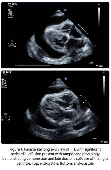
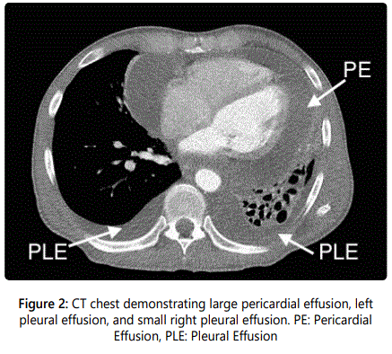
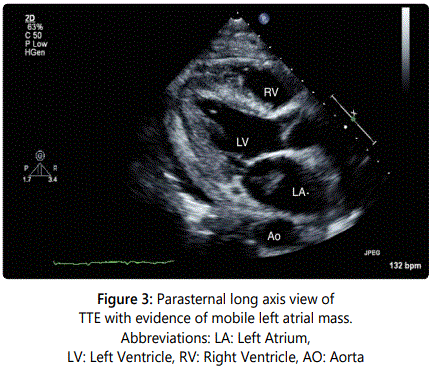
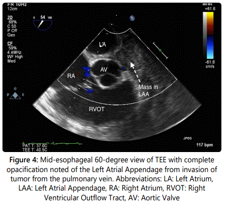
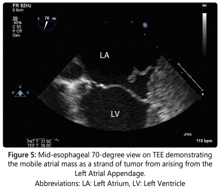
A 54-year-old gentleman with recently diagnosed Stage IIIB squamous cell carcinoma of the lung with metastases to the lymph nodes had a transthoracic echocardiograph (TTE) performed for evaluation of progressive dyspnea on exertion. A large pericardial effusion causing tamponade physiology was noted on TTE and the Computerized Tomography (CT) image of the thorax (Figures 1 and 2). Following a pericardi ocentesis, a repeat TTE was performed which showed the presence of a left atrial mass (Figure 3). The mass was initially reported as a 1.3x0.5 cm pedunculated mobile left atrial mass arising from the left atrial appendage prolapsing in the mitral orifice in diastole before further assessment with a transesophageal echocardiogram (TEE) was performed. The TEE demonstrateddirect invasion of the mass from the left upper pulmonary vein into the left atrial appendage with minimal residual flow of that pulmonary vein and complete opacification of the left atrial appendage as well as invasion of the superior vena cava (Figure 4). The mobile mass in the left atrium was a strand of tumor that extended from the edge of the left atrial appendage (Figures 5 and 6).
Comment
Metastases to heart are significantly more common than primary cardiac tumors with an approximate 20-40x the prevalence of that of primary tumors [1,2]. While historically a post-mortem finding, cardiac metastases are becoming increasingly diagnosed in living individuals with advances in imaging modalities including echocardiography (TTE and TEE), CT, MRI, and Positron Emission Tomography [3,4]. Here, we describe a rare example of direct invasion from squamous cell carcinoma of the lung of a mobile mass in the left atrium on TTE that was better visualized with TEE.
The patient underwent palliative radiation therapy before transitioning to hospice care.
Conflicts of Interest: The author reports no conflict of interest.
References
- Strauss BL, Matthews MJ, Cohen MH, et al. Cardiac metastases in lung cancer. Chest. 1977; 71(5): 607-611. http://dx.doi.org/10.1378/chest.71.5.607
- Sosinska-Mielcarek K, Senkus-Konefka E, Jassem J. Cardiac involvement at presentation of non-small-cell lung cancer. J Clin Oncol. 2008; 26(6): 1010-1011. doi: 10.1200/JCO.2007.14.9328
- Verma V, Talmon GA, Zhen WK. Intracardiac Metastasis From Non-Small Cell Lung Cancer. Front Oncol. 2015; 5: 168. doi: 10.3389/fonc.2015.00168
