International Conference on Vaccines
Feb 20-22, 2017 | Baltimore, USA
Synthesis and scanning electron microscopy investigation of polyvinyl pyrrolidone / silver composite nanofibers for application in biomedicine
1Department of Nanoscience and Technology, Bharathiar University, Tamilnadu, India
2National Centre for Nanosciences & Nanotechnology, University of Mumbai, India
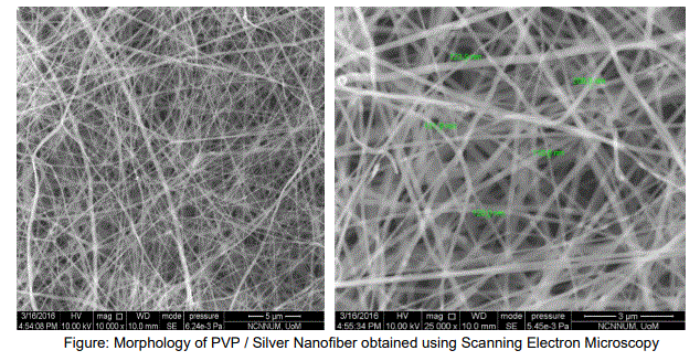
The electrospinning technique attracted much attention recently to the research due to the ability of converting materials to order of few nanometers with large surface areas, ease of functionalization for various purposes and superior mechanical properties. Also, the possibility of large scale productions combined with the simplicity of the process makes this technique very attractive for many different applications. Biomedical field is one of the important application areas among others utilizing the technique of electrospinning is used to produce fibers in the nanometer range by stretching a polymeric jet using electric field of high magnitude. Electrospinning leads to the formation of continuous fibers ranging from 100nm to 500nm. The ultra-fine fibers produced by electro spinning are expected to have two main properties, a very high surface to volume ratio, and a relatively defect free structure at the molecular level. Composites in the form of nanofibers were formed via electrospinning technique. Different ratios and different parameters pvp / silver composite nanofibers prepared in electrospinning. The prepared composite nanofibers were characterized using several techniques such as Scanning Electron Microscopy (SEM), X-ray Diffraction (XRD). It is anticipated that this PVP / silver nanofiber can be used in various applications such as clinical wound dressing, antimicrobial activity, bio adhesive, biofilm, and the coating of biomedical materials, etc. The morphology of PVP / silver nanofiber obtained using Scanning Electron Microscopy is show below.


