
Research Article
Functional Endonasal Sinus Surgery in Newborns and small children-25 Years' Experience
1Clinic of Pediatric Otorinolaryngology, University Hospital Brno, Jihlavska 20, 625 00 Brno, Czech Republic
2Faculty of Medicine, Masaryk University, Kamenice 5, 625 00 Brno, Czech Republic
*Corresponding author: Michaela Máchalova, Clinic of Pediatric Otorinolaryngology, University Hospital Brno, Jihlavská 20, 625 00 Brno, Czech Republic, Tel.: +420 532 234 440, E-mail: michaela.machalova@centrum.cz
Received: September 20, 2017 Accepted: October 26, 2017 Published: November 2, 2017
Citation: Šlapák I, Máchalová M, Urik M, Forstová G. Functional Endonasal Sinus Surgery in Newborns and Small Children-25 years' experience. Madridge J Otorhinolaryngol. 2017; 2(1): 26-30. doi: 10.18689/mjol-1000105
Copyright: © 2017 The Author(s). This work is licensed under a Creative Commons Attribution 4.0 International License, which permits unrestricted use, distribution, and reproduction in any medium, provided the original work is properly cited.
Abstract
The most frequent indication for endoscopic endonasal surgery in newborn children (0-1 month) is bilateral choanal atresia. In bilateral atresia, severe early respiratory problems are common and endotracheal intubation is often necessary. The second most frequent indication for FESS in newborns is congenital disorders of lacrimal passages.
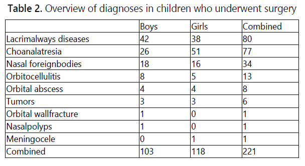
A sample of patients, who underwent functional endonasal sinus surgery from 1991 to 2015 at the Clinic of Paediatric Otorhinolaryngology, Masaryk University and Faculty Hospital Brno, was subjected to retrospective analysis in order to evaluate outcomes of this method. The nasal membrane was anesthetized preoperatively by inserting nasal tampons with the solution of Epinephrine 1:1000 or Nafazoline 0.05%. Rigid optics with diameters of 4 mm and 2.7 mm were used as standard. After the operation, an anterior greasy nasal packing was applied into the nasal cavity to prevent bleeding, if necessary. In the mentioned period, 1282 children (0-19 years of age) were operated at our clinic using the FESS method, of these were 750 boys (58 %) and 532 girls (42 %).From this sample, the group of youngest children aged 0-5 was examined in detail. In the group 221 children were included, of them 57 were newborns aged 0-1 month and 164 were children aged from 1 month to 5 years. The most frequent indication for the functional endonasal surgery in newborns was bilateral choanal atresia - 49 cases. Girls (32 cases) were slightly more numerous than boys (17 cases). The smallest child undergoing the surgery fort his diagnosis was born prematurely and weighed 1500 g. Children suffering from the bilateral choanal atresia were intubated using orotracheal tube upon the birth and operated on day 3 - 5 after their birth. The bony plate in the choana was removed and a part of the nasal septum was resected during the operation. The second most frequent indication in newborns was the congenital obstruction of lacrimal passages - 8 cases (4 boys and 4 girls). Those were mainly the cases of postsacal. In the group of infants and small children, the functional endonasal sinus surgery was most frequently used for the surgical treatment of obstructed lacrimal passages - in 72 cases. Using the Thulium laser for DCRS is particularly favourable with breast-fed children because in the case of bilateral defect it is not necessary to apply the nasal tamponade, which limits breathing of the child during feeding. Surgery for choanal atresia was performed on 28 patients in this group: bilateral in 8 cases and unilateral in 20 cases. Other indications included the removal of an acute stuck foreign body in a noncooperative patient (22 cases) and the extraction of a chronic foreign body, which has been often already covered in the granulation tissue. Cases of complicated inflammations of paranasal sinuses were also treated by FESS in this group, when the previous conservative therapy with antibiotics had failed. In 13 cases, unilateral ethmoidectomy and release of pathological content were carried out upon the finding of orbitocellulitis not responding to the antibiotic therapy. In 8 cases, an orbital subperiostalabscess was operated via ethmoidectomy. Probatory excision of a tumour in the area of nose, nasopharynx or paranasal sinuses was performed in 6 cases. Endoscopic endonasal approach is good for visualisation and targeted biopsy in order to obtain the material for the histopathological examination of a suspected malignant neoplasm. The most frequently occurring malignant tumour in the sample was rhabdomyosarcoma others were non-Hodgkin lymphoma and Ewing sarcoma. Uncommon indications for FESS in our sample included meningocele, orbital wall fracture and nasal polyps in 5 year old child suffering from cystic fibrosis. For detailed overview of diagnoses.
Keywords: Surgery; Endonasal Sinus; Atresia; Congenital disorders; Bleeding and Orbitocellulitis
Introduction
Methods of functional endonasal sinus surgery are used for diagnostic and surgical procedures in all age categories [1-3]. Technical development of used optics and endonasal instrumentation made it possible to use this technique even in newborns and infants, at whom the operating space is significantly smaller than in adolescents and adults [4].
In newborns, only ethmodial sinuses are developed. In the period of growth up to an age of approximately 10-12 years, other paranasal sinuses develop in a proces, which is called skull pneumatisation. For this reason, the range of FESS application in the category of youngest children is different from that used in older children and adults [5].
In the youngest children, this technique is used for diagnostics and therapy of congenital disorders in the area of nose and nasopharynx and for the surgical treatment of obstructed nasolacrimal duct.
The most frequent indication for endoscopic endonasal surgery in newborn children (0-1 month) is bilateral choanal atresia. Congenital choanal atresia is the most common disorder in the nasal area; incidence rate is 1 per 7-8000 newborns and it affects girls more often than boys (2:1). Choanal atresia can be both uni- and bilateral. In bilateral atresia, severe early respiratory problems are common and endotracheal intubation is often necessary. Children undergo surgery usually during the first week after birth [6,7].
The second most frequent indication for FESS in newborns are congenital disorders of lacrimal passages. Dacryocystorhinostomy is the most common procedure in infants and toddlers, when the conservative therapy fails [8].
The period between 2 and 4 years is the age, in which children explore not only the surrounding environment but also their own bodies, finding places where various things can be hidden. Foreign bodies in the nose are the most frequently diagnosed problem in this age category [9]. Endoscopic endonasal instrumentation can be also used in their diagnostics and extraction. In the period between year 1 and 5 of age, complications of paranasal sinuses inflammation may occur such as acute orbitocellulitis and sporadically also a serious complication - orbital abscess, which - if not treated surgically in due time - might result in the impairment of patient's vision [10]. This age group may also be affected by malignancies in the area of nose, paranasal sinuses and nasopharynx.
Material and Methods
A sample of patients, who underwent functional endonasal sinus surgery from 1991 to 2015 at the Clinic of Pediatric Otorhinolaryngology, Masaryk University and Faculty Hospital Brno, was subjected to retrospective analysis in order to evaluate outcomes of this method. The hospital provides healthcare for children and young people between 0 and 19 years of age.
The patients were operated in general anaesthesia with air passages secured either by orotracheal intubation or laryngeal mask. The nasal membrane was anesthesized preoperatively by inserting nasal tampons with the solution of Epinephrine 1:1000 or Nafazoline 0.05%. The tampons were put into the nasal cavity 10-15 minutes before the beginning of the operation. During the operation, patients were positioned on the back with their head slightly bent forward and the surgeon was standing at the patient's right side. Rigid optics with diameters of 4 mm and 2.7 mm were used as standard. The optical equipment was fixed in a holder with a washing system. Standard endoscopic endonasal surgery instruments were used for the operation. Since 2012, Thulium laser was also used for surgeries, the main benefit of this device is the significant suppression of bleeding. After the operation, an anterior greasy nasal packing was applied into the nasal cavity to prevent bleeding. In the smallest children, it is much better than epistaxis catheter because of its size, which makes it more difficult for them to extract. The nasal packing was removed gradually within day 2 and 4 after the surgery.
In the mentioned period, 1282 children (0-19 years of age) were operated at our clinic using the FESS method, of these were 750 boys (58 %) and 532 girls (42 %).
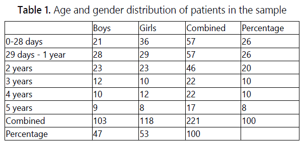
From this sample, the group of youngest children aged 0-5 was examined in detail. In the group 221 children were included, of them 57 were newborns aged 0-1 month and 164 were children aged from 1 month to 5 years . For the detail overview, see Tab. 1.
Results
3.1. Category of newborns (0-28 days)
The most frequent indication for the functional endonasal surgery in newborns was bilateral choanal atresia - 49 cases. Girls (32 cases) were slightly more numerous than boys (17 cases). The smallest child undergoing the surgery fort his diagnosis was born prematurely and weighed 1500 g. Children suffering from the bilateral choanal atresia were intubated using orotracheal tube upon the birth and operated on day 3 - 5 after their birth. The bony plate in the choana was removed and a part of the nasal septum was resected during the operation, in order to prevent possible stenosis or repeated obstruction of the choana. Neither stents nor the mitomycine solution were used on the mucous membranes. Greasy anterior nasal packing was applied to prevent or stop bleeding. It was removed on the second day after the surgery. Subsequently, the mucous membrane of choana was treated under endoscopic control with crusts and fibrin depositions being removed. Nasal mucosa was moistened with the saline solution or sea water solutions and panthenol oilments were applied locally.
The second most frequent indication in newborns was the congenital obstruction of lacrimal passages - 8 cases (4 boys and 4 girls). Those were mainly the cases of postsacal stenosis, in which suprasacal lacrimal ways are developed including puncta lacrimalia. In cases when the puncta or one of them is not developed, the surgical treatment is complicated. When only one lacrimal punctum was present, a silastic tubing implant was inserted into the lacrimal passages and fixed to the eyelid. Creating an arteficial lacrimal point by simple perforation is not recommended, because after the extraction of the tube it tends to regress. Preoperative treatment is similar as in the other endonasal surgeries.
The FESS instrumentation was also used for diagnosing suspected pathologies in the nasal cavity or nasopharynx, mostly in multiple congenital disorders.
3.2. Category of infants and small children (29 days - 5 years of age)
In the group of infants and small children, the functional endonasal sinus surgery was most frequently used for the surgical treatment of obstructed lacrimal passages - in 72 cases. During the operation, mucosa in front of concha nasalis media was separated instrumentally and the lacrimal bone was denuded. In the last years of the study period (2012- 2015), the Thulium laser device was used for separating of the mucous membrane, which helped to control bleeding. The partial ablation of the mucous membrane and lacrimal bone was followed by lacrimal sac opening. Subsequently, a tube with a silicone fibre was inserted through the lower and upper puncta lacrimalia, which was led under endoscopic control into the nose and knotted. The silicone fiber knot acts as a foreign body, and the organism reacts to it by creating cicatricial tissue around the fibre. The process results in the creation of an arteficial duct, which serves to drain tears physiologically into the nasal cavity. When dacryocystorhinostomy (DCRS) is performed in this way, the fibre is left inside for approximately 6 months and then it is removed, usually at an outpatient check-up.
Using the Thulium laser for DCRS is particularly favourable with breast-fed children because in the case of bilateral defect it is not necessary to apply the nasal tamponade, which limits breathing of the child during feeding. Both peroperative and postoperative bleeding is almost absent when the Thulium laser is used.
Surgery for choanal atresia was performed on 28 patients in this group: bilateral in 8 cases and unilateral in 20 cases. Other indications included the removal of an acute stuck foreign body in a noncooperative patient (22 cases) and the extraction of a chronic foreign body, which has been often already covered in the granulation tissue (12 cases). Cases of complicated inflammations of paranasal sinuses were also treated by FESS in this group, when the previous conservative therapy with antibiotics had failed. In 13 cases, unilateral ethmoidectomy and release of pathological content were carried out upon the finding of orbitocellulitis not responding to the antibiotic therapy. In 8 cases, an orbital subperiostal abcess was operated via ethmoidectomy. Probatory excision of a tumor in the area of nose, nasopharynx or paranasal sinuses was performed in 6 cases. Endoscopic endonasal approach is good for visualisation and targeted biopsy in order to obtain the material for the histopathological examination of a suspected malignant neoplasm. The most frequently occurring malignant tumor in the sample was rhabdomyosarcoma others were non-Hodgkin lymphoma and Ewing sarcoma. Uncommon indications for FESS in our sample included meningocele, orbital wall fracture and nasal polyps in 5 year old child suffering from cystic fibrosis. For detailed overview of diagnoses, see Tab. 2.
Reoperation of choanal atresia was necessary in six cases in the group of newborns. In all of the cases, the reason was a repeated stenosis of choanae in children with multiple congenital defects in the craniofacial area. In the group of older children (1 month-5 years), the DCRS reoperation was performed in three cases due to the repeated epiphora and blockage of lacrimal passages.
Serious perioperative complications such as intracranial space penetration or significant bleeding were not recorded.
Discussion
In the last decades, there is an obvious shift in indications for functional endonasal sinus surgery to the lowest age categories including newborns. A new opportunity to use modern devices and instruments such as thin optics, shaver and laser comes with the technical development. Another reason for this trend is the expansion of knowledge and skills in endoscopic endonasal surgery [4,5].
In order to acquire necessary skills, it is favorable to start operating adults or adolescents first since anatomical structures are already developed, the operating field is larger, clearer, and the orientation is thus easier [1,3]. In the youngest children, anatomic ratios in the area of nose and paranasal sinuses are different and require experience, a low-impact operating technique and skills of the surgeon.
Results from our group of patients confirm that the most frequent indication for using the functional endonasal sinus surgery in newborns are congenital anomalies e.g. bilateral choanal atresia or defects of lacrimal passages [6,7,8]. Operation techniques for the surgical treatment of the congenital bilateral choanal atresia are identical at most pediatric ENT departments. Reduction in the length of surgery and minimum impact on nasal and paranasal tissues are indisputable advantages of the endoscopic approach as compared with the conventional technique [11,12].
Technical difficulties may occur in premature born children with a low birth weight and often suffering from multiple congenital defects, particularly in the orofacial area. Congenital bilateral choanal atresia is common in children suffering from Treacher Collins syndrome and CHARGE syndrome (the letters stand for: coloboma of the eye, heart defects, atresia of the nasal choanae, retardation of growth and/or development, genital and/or urinary abnormalities, and ear abnormalities) [13,14]. Using the endonasal endoscopic approach in children aged from 1 month to 5 years is most common in the case of obstructed lacrimal apparatus. Congenital anomalies or postinfectious stenoses are other frequent indications [8,15,16].
When the conservative therapy fails, opening of tear drainage ways by DCRS is a procedure, which does not significantly burden the patient and has a very good long-term effect [17,18]. Diagnostic rhinoscopy and rhinoepipharyngoscopy are often used for diagnosing nasal obstructions and in the cases of suspected pathological processes in the nasal cavity and nasopharynx [19,20].
Foreign bodies in the nasal cavity mostly occur in children between 2 and 4 years old and represent the most common foreign bodies in the ORL area. In most cases, they are acute foreign bodies, when the body is present in the nasal cavity for several hours or days. The foreign bodies can usually be easily removed during outpatient treatment, with the exception of stuck foreign bodies in noncooperative patients. Chronic foreign bodies as a cause of nasal blockage are diagnosed less often. From 2000 to 2008, 1421 foreign bodies at the Clinic of Pediatric ORL in Brno were removed, of which 704 from the nasal cavity. Balls, beads and toy parts were the most common objects, while batteries were the most dangerous ones. When the ambulatory extraction fails, it is necessary to perform extraction in general anaesthesia using endoscopic endonasal instruments [9].
The least common cases in the group of small children are chronic inflammations of the paranasal sinuses. More frequent are complications of acute inflammations of paranasal sinuses, especially in the area of ethmoidal cells and maxillary sinuses. The most common of these complications is acute orbital cellulitis. If the conservative therapy with antibiotics fails in orbitocellulitis or orbital abscess is revealed by diagnostic imaging, it is necessary to perform a surgery via endonasal ethmoidectomy and abscess evacuation [10,21]. Formerly it was necessary to operate patients with these complications using the external access. Nowadays, when using the FESS method, it is no longer necessary, and patients can benefit from the faster recovery and shorter hospital stay. Also the skin in the facial area is not damaged by the endoscopic approach [22].
Conclusions
Indications for endoscopic endonasal surgery in newborns are congenital anomalies of choanae and lacrimal apparatus. In children aged from 1 month to 5 years, the most common surgical procedure using the endoscopic approach is the release of obstructed lacrimal passages and choanal atresia. Operations of inflammatory complications in paranasal sinuses and orbits are less frequent. Nasal endoscopy is also used for diagnosing pathological conditions in the nasal cavity and nasopharynx. Prerequisites of success are lowimpact operation technique and adequate instrumentation.
Conflicts of Interest: The authors declare no conflicts of interest with this submission.
References
- Lazar RH, Younis RT, Long TE. Functional endonasal sinus surgery in adults and children. Laryngoscope. 1993; 103(1): 1-5. doi: 10.1288/00005537- 199301000-00001
- Stammberger H, Posawetz W. Functional endoscopic sinus surgery. Eur Arch Otorhinolaryngol. 1990; 247: 63-76. doi: 10.1007/BF00183169
- Hybášek I, Vokurka J. Functional endonasal surgery. conception a indications. Otorinolaryn Foniatr. 1994; 43: 60-62.
- Chang PH, Lee LA, Huang CC, Lai, Lee TJ. Functional Endoscopic Sinus Surgery in Children Using a Limited Approach. Arch Otolaryngol Head Neck Surg. 2004; 130(9): 1033-1036. doi: 10.1001/archotol.130.9.1033
- Gross CW, Guruchiarl MJ, Lazar RH, Long TE. Functional endonasal sinus surgery (FESS) in the pediatric age group. Laryngoscope. 1989; 99: 272- 275. doi: 10.1288/00005537-198903000-00007
- Newman JR, Harmon P, Shirley WP, Hill JS, Woolley AL, Wiatrak BJ. Operative management of choanal atresia: a 15-year experience. JAMA Otolaryngol Head Neck Surg. 2013; 139(1): 71-5. doi: 10.1001/ jamaoto.2013.1111
- Zuckerman JD, Zapata S, Sobol SE. Single-stage choanal atresia repair in the neonate. Arch Otolaryngol Head Neck Surg. 2008; 134(10): 1090-3. doi: 10.1001/archotol.134.10.1090
- Cunningham MJ. Endoscopic management of pediatric nasolacrimal anomalies. Otolaryngol Clin North Am. 2006; 39(5): 1059-74. doi: 10.1016/j.otc.2006.07.004
- Slapak I, et al. Non Food Foreign Body injurie. Int J Pediatr Otorhinolaryngol. 2012; 76(1): 26-32. doi: 10.1016/j.ijporl.2012.02.006Oxford LE, McClay J. Medical and surgical management of subperiostal orbital abscess secondary to acute sinusitis in children. Int J Pediatr Otorinolaryngol. 2006; 70(11): 1853-61. doi: 10.1016/j.ijporl.2006.05.012Gosepath J, Santamaria VE, Lippert BM, Mann WJ. Forty-one cases of congenital choanal atresia over 26 years--retrospective analysis of outcome and technique. Rhinology. 2007; 45(2): 158-63.Teissier N, Kaguelidou F, Couloigner V, François M, Den Abbeele TV. Predictive factors for success after transnasal endoscopic treatment of choanal atresia. Arch Otolaryngol Head Neck Surg. 2008; 134(1): 57-61. doi: 10.1001/archoto.2007.20Den Abbeele TV, Francois M, Narcy P. Transnasal endoscopic treatmento choanal atresia without prolonged stenting. Arch Otolaryngol Head Neck Surg. 2002; 128(8): 936-940.Keller JL, Kacker A. Choanal atresia, Charge assotiation and congenital nasal stenosis. Otolaryngologic Clinics of North America. 2000; 33(6): 1343-1351. doi: 10.1016/S0030-6665(05)70285-1Kominek P, Cervenka S, Pniak T, Zelenik K, Tomaskova H, Matousek P. Revison endonasal dacryocystorhinostomies: analysis of 44 procedures. Rhinology. 2011; 49(3): 375-80. doi: 0.4193/Rhino10.293Kouri AS, Tsakanikos M, Linardos E, Nikolaidou G, Psarommatis I. Results of endoscopic assisted probing for congenital nasolacrimal duct obstruction in older children. Int J Pediatr Otorhinolaryngol. 2008; 72(6): 891-6. doi: 10.1016/j.ijporl.2008.02.024Gioacchini FM, Cinfelli MA, Kaleci S, Re M. Theoutcomes of endoscopic dacryocystorhinostomy in children: A systemic review. Int Pediatr Otorhinolaryngol. 2015; 79(7): 947- 52. doi: 10.1016/j.ijporl.2015.04.023Lee S, Yen MT. Laser - assisted dacryocystorhinostomy: a viable treatment option? Current Opinion in Ophthalmology. 2011; 22(5): 413-18. doi: 10.1097/ICU.0b013e32834994c8Chigurupati R, Alfatooni A, Myall RWT, Hawkins D, Oda D. Orofacial rhabdomyosarcoma in neonates and young children: a review of literature and management of four cases. Oral Oncol. 2002; 38(5): 508-15. doi: 10.1016/S1368-8375(01)00087-2Hicks J, Flaitz C. Rhabdomyosarcoma of the head and neck in children. Oral Oncology. 2002; 38(5): 450-59. doi: 10.1016/S1368-8375(01)00105-1Nageswaran S, Woods ChR, Benjamin DK Jr, Givner LB, Shetty AK. Orbital cellulitis in children. Pediatric Infect Disease Journal. 2006; 25(8): 695-99.Deutsch E, Eilon A, Hevron I, Hurvitz H, Blinder G. Functional endoscopic sinus surgery of orbital subperiostal abscess in children. Int J Pediatr Otorinolaryngol. 1996; 34: 181-190. doi: 10.1016/0165-5876(95)01253-2
