
Research Article
Middle Ear Risk Index [MERI] as Prognostic Factor in Tympanomastoidectomy with Tympanoplasty
1Senior Resident, Department of Otorhinolaryngology, J.N. Medical College, Aligarh Muslim University, Aligarh 202002, India
2Professor, Department of Otorhinolaryngology, J.N. Medical College, Aligarh Muslim University, Aligarh 202002, India
*Corresponding author: Aftab Ahmed, Senior Resident, Department of Otorhinolaryngology, J.N. Medical College, Aligarh Muslim University, Aligarh 202002, India, Tel: +918791520112, E-mail: aftab2k3@gmail.com
Received: March 11, 2016 Accepted: May 12, 2016 Published: May 17, 2016
Citation: Ahmed A, Sharma SC. Middle Ear Risk Index [MERI] as Prognostic Factor in Tympanomastoidectomy with Tympanoplasty. Madridge J Otorhinolaryngol. 2016; 1(1): 15-22. doi: 10.18689/mjol-1000103
Copyright: © 2016 The Author(s). This work is licensed under a Creative Commons Attribution 4.0 International License, which permits unrestricted use, distribution, and reproduction in any medium, provided the original work is properly cited.
Abstract
Aims: To evaluate a group of patients undergoing surgery for chronic otitis media with reference to the prognostic value of middle ear risk index and other factors in predicting the anatomical and functional outcome of tympanomastoidectomy with tympanoplasty.
Study Design: Prospective study.
Setting: Tertiary referral hospital.
Subjects: The study comprised of 90 patients suffering from chronic otitis media with or without cholesteatoma. Patients attending the Otorhinolaryngology out patients department were considered for this study.
Methods: The patients underwent tympanomastoidectomy with tympanoplasty in which mastoidectomy performed was of either canal wall up or canal wall down technique. In cholesteatoma surgery, whenever possible a canal wall up procedure was performed. Myringoplasty was done using autologous temporalis fascia graft by underlay technique. Ossiculoplasty was performed by autologous incus interposition, partial ossicular prosthesis or total ossicular prosthesis, depending upon the status of the remanant ossicles. Patients were followed up for a period of 6 months. Middle ear risk index [MERI] and other factors were evaluated for their outcome predictive role in patients undergoing tympanomastoidectomy with tympanoplasty.
Results: Outcomes were evaluated in terms of tympanic membrane graft uptake and post operative mean audiological gain. The presence of cholesteatoma, granulation tissue, mucosal polyp, ossicular necrosis, patency of Eustachian tube, mastoidectomy technique and attempt of surgery affect tympanic membrane graft uptake as well as mean audiological gain. The size of the perforation has no effect on both outcomes,
whereas presence of tympanosclerotic patch has a significant effect only on mean audiological gain. The Middle ear risk index was also found to be significant predictor of the outcome of surgery. The patients with mild MERI scores had significantly better prognosis than the patients with sever MERI scores.
Conclusion: The Anatomical and Functional outcome of tympanomastoidectomy with tympanoplasty is diversely affected by the pathological and technical factors associated with disease and its management. A better understanding of these factors is helpful for better prognostication of the factors affecting the disease and in planning the surgical procedure.
Keywords: Middle ear risk index; Tympanomastoidectomy; Tympanoplasty; Chronic otitis media; Prognostic factors; Canal wall up; Canal wall down
Abbreviations: MERI: Middle Ear Risk Index; COM: Chronic Otitis Media; SPITE: Surgical,
Prosthetic, Infection, Tissue and Eustachian Tube; CWU: Canal Wall Up; CWD: Canal Wall Down
Introduction
The primary objective of surgery for chronic otitis media
(COM) is to eradicate the disease and to make the ear safe and dry. The incidence of the ears becoming dry after surgery and the ears not having recurrent or residual cholesteatoma ranges between 70 to 90 percent in various large clinical trials
[1]. A second objective of surgery for COM is to restore hearing to serviceable levels by means of tympanoplasty.
There has been difference in opinion about the staging of the surgical procedure for COM. Some studies supported the single stage surgery for both elimination of disease and tympanoplasty [2,3]. Whereas others advocate two stage procedure for achieving the different objectives [4,5].
Tympanomastoidectomy is the procedure for removal of disease from middle ear cleft done either as open or closed cavity procedure, and tympanoplasty is the procedure for reconstruction of the middle ear. The success of tympanomastoidectomy with tympanoplasty is dependent not only upon the surgical principle but also on the pathological factors associated with disease. Although there is huge literature present about the techniques of tympanomastoidectomy with tympanoplasty but the data about factors affecting the outcome is limited. The pathologic condition of the middle ear as a predictor of outcome has been confusing issue in the literature [6-8]. The decision for single or two stage procedure for COM can be made depending upon the pathological factors associated with disease. For this purpose a grading system has been devised,
known as Middle ear risk index (MERI).
Middle Ear Risk Index (MERI) of a patient suffering from the COM is a numerical grading to stratify the severerity of disease. MERI is determined by assigning a specific value for each risk factor, and these values are added to get the MERI score. The risk factors include Belluci criteria to assess the degree of otorrhoea, Austin/Kartush criteria for ossicular status, presence of perforation, cholesteatoma middle ear granulations/effusion and history of previous surgery. The suggested risk categories can be derived from MERI as follows:
MERI 0 = Normal; MERI 1-3 = Mild disease; MERI 4-6 =
Moderate disease; MERI 7-12 = Severe disease [9].
In the developing countries the cost of surgery and absence from the work are major restrains for two stage surgical procedure. If we can predict the outcome of the surgical procedure depending upon the pathologic condition of the middle ear, the cost effectiveness of the surgery can be improved and this will also improve the patients compliance.
The pathological factors affecting the surgical outcome of COM has been highlighted by different authors time by time but factors have been analyzed separately and each study has concentrated only on one factor at a time [10-12]. There is only one study combining these factors, the surgical, prosthetic,
infection, tissue and Eustachian tube (SPITE) method which is an exception [6]. This makes it imperative for studying various pathological factors of middle ear disease simultaneously and to provide guidelines for their management.
The objective of this study is, to evaluate a group of patients undergoing tympanomastoidectomy with tympanoplasty with reference to the prognostic significance of Middle ear risk index
(MERI) and other factors in predicting the anatomical and functional outcome of the surgery.
Materials & Methods
This study has been carried out in the Department of Otorhinolaryngology, Jawaharlal Nehru Medical College,
Aligarh Muslim University, Aligarh, India from January 2012 to November 2015. The study comprised of 90 patients suffering from chronic otitis media with or without cholesteatoma. The age of patients ranged from 7 to 46 years and male to female sex ratio of 1.3:1. The patients were subjected to detailed history, general as well as systemic examination, which includes clinical examination of the ear, nose, paranasal sinuses, larynx and pharynx. The complete otological evaluation has been done to assess the exact nature and extent of disease, presence or absence of tympanosclerosis,
cholesteatoma, granulation tissue, mucosal polyp and ossicular chain status. Eustachian tube function was evaluated by valsalva test and Eustachian tube cathetarisation. Pure tone audiometry was done for audiological evaluation and readings were taken at 500, 1000, 2000 and 4000 Hz and AB gap was calculated by taking average of above four frequencies.
Complete laboratory evaluation was done and radiological evaluation was done if needed.
The size of the perforation was graded as small (less than
50%), medium (50-75%) and large (> 75%) of the total tympanic membrane area. The size of the perforation was compared for its effect on the outcome of surgery.
Mastoidectomy performed was of either canal wall up (CWU)
or canal wall down (CWD) technique. In cholesteatoma surgery, whenever possible a canal wall up procedure was performed. Canal wall down technique was done in cases having extensive disease, erosion of the external auditory meatus, or revision surgery for extensive recurrent disease.
CWU procedure was done in 67 patients and CWD procedure was done in 23 patients. Myringoplasty was performed in all the 90 patients using autologous temporalis fascia grafts by underlay technique. The ossicular reconstruction technique was chosen based on the available ossicular remnants after the complete removal of diseased tissues. Ossiculoplasty was performed in 57 patients, autologous incus interposition,
partial ossicular prosthesis and total ossicular prosthesis were used depending upon the status of middle ear ossicles.
In our study of 90 patients, 68 patients were having tympanic membrane perforation and 22 patients were having retraction pockets. Out of 68 patients, 8 patients were having anterior perforation, 24 patients were having central perforation, 19 patients were having posterior perforation and 17 patients had marginal or attic perforation. The number of smokers in our study was 18 patients out of 90 patients. The age of the patients in our study ranged from 7 years to 46
years. The number of patients in the first decade of life were
6, in the second decade of life were 28, in the third decade of life were 26, in the forth decade of life were 21 and in the fifth decade of life were 9. The location of the cholesteatoma in the patients included in our study was in the epitympanum in 15
patients and in the posterior-superior region of the middle ear in 22 patients. In our study the size of the cholesteatoma was not measured. In our study ossiculoplasty was performed by autologous incus interposition in 27 patients, partial ossicular prosthesis was used in 20 patients, and total ossicular prosthesis was used in 10 patients. These variables have been summarised in a table under the results section. All the patients included in the present study were operated by the same surgeon and the surgical technique remained the same throughout the period of study.
The middle ear risk index was calculated for each patient and mean audiological gain in group of the patients having mild, moderate and severe MERI were compared and their statistical significance was studied. Statistical significance of other factors were also evaluated.
Data Collection Technique and Tools
Inclusion Criteria:
1. Patients suffering from unilateral or bilateral chronic
otitis media with or without cholesteatoma.
2. Patients between 7 to 46 years of age.
3. Patients with adequate cochlear reserve.
Exclusion Criteria:
1. Patients with sensorineural deafness.
2. Patients found to have systemic disease e.g. hypertension,
diabetes mellitus.
3. Patients with Adenotonsillitis, cleft palate and nasal polyp.
4. Patients with intracranial complications of chronic otitis media.
Patients were followed up for a period of about 6 months.
As most of the patients in the present study were from rural area, they do not return for follow up once they have improvement after the surgery. In our study of 90 patients,
only 81 patients came for the follow up at the 6 months after the surgery, 9 patients were lost for follow up. The anatomical and functional outcome of tympanomastoidectomy with tympanoplasty was evaluated in terms of tympanic membrane graft uptake at 3 months and mean audiological gain taking into consideration the average of the readings at 500, 1000,
2000 and 4000 Hz after 3 and 6 months of surgery. Mean audiological gain for different categories of MERI was compared at 3 months and 6 months
Statistical Analysis
Data was analyzed by using SPSS 20. Chi- square, unpaired t-test and one way analysis of variance (ANOVA) test was used for statistical comparison. Tukey's Honest Significant Difference post hoc test was used after ANOVA. P value of <0.05 was considered significant while p value <0.01 was considered highly significant.
Results
The present study has been carried out from January 2012
to November 2015. The study comprised of 90 patients suffering from COM with or without cholesteatoma, of the age ranging between 7-46 years and of both the sexes.
In our study, out of 90 patients, 68 patients had tympanic membrane perforation and 22 retraction pocket. Out of 68
patients, 21 had small size, 35 medium size and 12 patients had large size perforations. Middle ear pathological conditions such as tympanosclerosis, cholesteatoma, granulation tissue and mucosal polyps were also evaluated as prognostic factors.
In our study tympanosclerosis was present in 18 patients,
cholesteatoma was present in 37 patients, granulation tissue was present in 28 patients and mucosal polyp was seen in 13
out of total 90 patients. Eustachian tube function was evaluated by Valsalva test and Eustachian tube cathetarisation and was found to be patent in 48 patients and blocked in 42
patients. The technical factors of surgery such as mastoidectomy technique and attempt of the surgery were also taken into consideration. CWU mastoidectomy was done in 67 patients and CWD mastoidectomy was performed in 23
patients. Revision surgery was done in 12 cases whereas in 78
patients it was primary surgery.
The variables such as site of perforation, age distribution of the patients and numbers of the ossicular prosthesis used
(autologous incus, PORP & TORP) in the study did not have significant effect on either tympanic membrane graft uptake or mean audiological gain after 3 months of surgery, whereas numbers of smokers in the study and location of the cholesteatoma had statistically significant effect on both tympanic membrane graft uptake and mean audiological gain after 3 months of surgery. The results of these variables are summarised in Table-1 and Table-2.
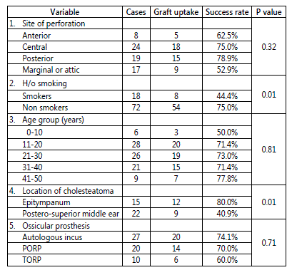
Table 1: Graft uptake after 3 months of surgery for certain variables of the patients included in the study.
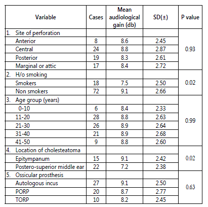
Table 2: Mean audiological gain after 3 months of surgery for certain variables of the patients included in the study.
The results in tympanomastoidectomy with tympanoplasty depend on variety of factors related to both the pathologic condition and the surgical technique. In our study the purpose was to evaluate whether these variables involved in the surgery for COM can predict the anatomical and functional outcome postoperatively. Surgical technique was not taken into account in our study, because the technique used was same throughout the period of study. The factors which contribute to middle ear risk index were studied for their prognostic value and other factors predicting the outcome of surgery were also evaluated.
The tympanic membrane graft uptakes were evaluated at
3 months postoperatively and mean audiological gain at 3
and 6 months postoperatively. Mean audiological gain in patients of different categories of MERI was compared at 3
and 6 months after surgery.
Tympanic Membrane Graft Uptake
Tympanic membrane graft uptake in tympanomastoidectomy with tympanoplasty was evaluated at 3 months post operatively. On otoscopic examination successful uptake of graft was taken only in those patients in whom no remnant of perforation was present in any of the quadrant of tympanic membrane. Graft uptakes for different variables studied are summarized in Table 3. Part-A of the table contains the variables included in MERI, whereas Part-B contains other factors analyzed.
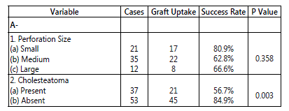
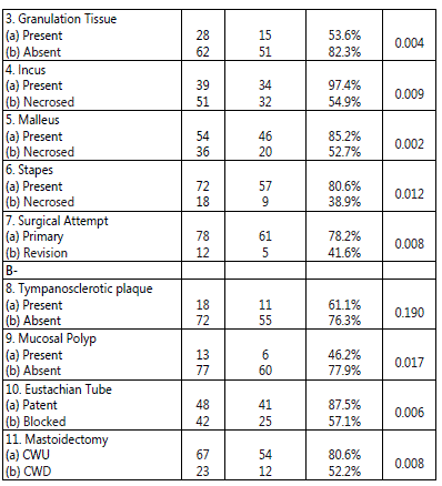
Table 3: Tympanic membrane graft uptake after 3 months of surgery.
Audiological Gain
Pure tone audiometry following tympanomastoid surgery was done at 3 and 6 months and audiological improvement
(taken as closure of air bone conduction gap) was measured in all patients undergoing the surgery for chronic otitis media.
AB gap (air - bone conduction gap) per case was calculated as mean of AB gap at four frequencies (500Hz, 1000Hz, 2000Hz
& 4000 Hz) pre operatively and post operatively.
Audiological gain was calculated for each patient by subtracting the post op AB gap from the pre op AB gap. The mean was calculated for each variable by dividing the sum of audiological gain in that group by the total number of cases in the same group.
We evaluated the mean audiological gain for different variables at 3 and 6 months and mean audiological gain for different categories of MERI at 3 and 6 months.
Pure Tone Audiometry Result at 3 months: In our study of
90 patients, pure tone audiometry was done 3 months postoperatively and audiological gain was evaluated. Audiological gain at 3 months for all the variables are summarized in Table 4. Part-A of the table contains the variables included in MERI, whereas Part-B contains other factors analyzed.
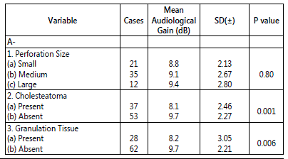
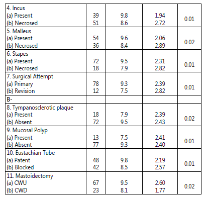
Table 4: Mean audiological gain after 3 months of surgery.
Pure Tone Audiometry Result at 6 months: In our study out of 90 patients only 81 patients attended the follow-up check after 6 months of surgery, 9 patients were lost for follow up. The audiological results at 6 months are summarized in Table 5. Part-A of the table contains the variables included in MERI, whereas Part-B contains other factors analyzed.
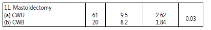
Table 5: Mean audiological gain after 6 months of surgery.
The tympanic membrane perforation size did not significantly affected the anatomical outcome i.e. graft uptake as well as functional outcome i.e. mean audiological gain at 3
and 6 months postoperatively.
The middle ear pathological conditions diversely affected both the outcomes. Tympanosclerosis did not significantly affected the graft uptake but it significantly affected the mean audiological gain at 3 and 6 months postoperatively. Whereas all other pathological conditions of the middle ear (cholesteatoma,
granulation tissue, mucosal polyp and ossicular necrosis) significantly affected both the graft uptake and mean audiological gain.
Pre operative Eustachian tube patency significantly affected the graft uptake as well as the mean audiological gain following tympanomastoidectomy with tympanoplasty. The technical factors, mastoidectomy technique (CWU or CWD) and attempt
(primary or revision) of the mastoidectomy, both significantly affected the graft uptake and mean audiological gain.
Mean audiological gain in terms of MERI in patients with dry ear at 3 months: The mean audiological gain in group of patients with mild, moderate and severe MERI at 3 months after surgery had difference which was statistically significant having a p-value of 0.01. On applying post hoc Tukey's Honest Significant Difference test between mild, moderate and severe MERI groups, it was found that there was statistically significant difference between mild and severe MERI patients with p-value of (P=0.01). The mean audiological gain for different categories of MERI at 3 months postoperatively are shown in Table 6.
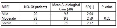
Table 6: Mean audiological gain in terms of MERI in patients withdry ear at 3 months.
Mean audiological gain in terms of MERI in patients with dry ear at 6 months: The mean audiological gain in group of patients with mild, moderate and severe MERI at 6 months after surgery had difference which was statistically significant having a p-value of 0.02. Post hoc Tukey's Honest Significant Difference test was applied to find the difference between mild, moderate and severe MERI groups. It was found that there was statistically significant difference between mild and severe MERI patients with p-value of (P=0.04). The mean audiological gain for different categories of MERI at 6 months postoperatively are shown in Table 7.
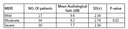
Table 7: Mean audiological gain in terms of MERI in patients with dry ear at 6 months.
Discussion
The principle of management of chronic otitis media is removal of diseased mucosa from the middle ear cleft and an attempt at restoration of hearing. The present study was conducted to assess the prognostic value of the various pathological and the technical factors associated with the COM on the outcome of the surgery. As stated earlier that the cost of the surgery and absence from the work are major restrains for the staged surgical procedure in the developing countries. The staging of the surgical procedure according the pathological condition of the middle ear will improve the outcome of the surgery and the compliance of the patients.
Middle ear risk index [MERI] is a numerical grading system to stratify the severity of the disease in the patients suffering from the COM. MERI can be used to decide the staging of the surgical procedure according to the severity of the disease.
MERI score was calculated for each patient. Mild, moderate and severe MERI groups were compared for outcome of surgery. MERI was found to be a predictor of outcome in the ear surgeries. The factors analyzed in the present study include perforation size, presence of tympanosclerosis, cholesteatoma,
granulation tissue, mucosal polyp, ossicular necrosis,
Eustachian tube patency, mastoidectomy technique and primary or revision surgery in COM patients. Some of the factors were constituents of the MERI whereas others were the additional factors analyzed. These factors were studied in COM patients undergoing tympanomastoidectomy with tympanoplasty for their effect on anatomical and functional outcome of the surgery, evaluated in terms of tympanic membrane graft uptake and audiological gain. We will here discuss the effect of various factors studied on the outcome of surgery after that we discuss the effect of MERI in predicting the outcome of surgery.
Tympanic Membrane Graft Uptake
Tympanic membrane perforation size was evaluated as determinant of graft uptake. Although lower success rates were observed for patients with larger tympanic membrane perforations, statistical analysis demonstrated no significant difference in surgical success rates between the various perforation size categories (p=0.35). Thus, on the basis of this study, perforation size was not predictive or determinant of successful tympanomastoidectomy with tympanoplasty. Our premise is on the basis that underlay technique of tympanoplasty with adequate graft size covers the membrane defect whether large or small. This result is similar to those of Wasson JD, et al.
[10], Pignataro L, et al. [11], Yung MW, et al. [13], Baloch MA,
et al. [14] and Vartiainen E, et al. [15] who have not given any plausible explanation for the conclusion.
In our study cholesteatoma was present in 37 patients. The success of graft uptake following tympanomastoidectomy with tympanoplasty was 56.7% in patients with cholesteatoma and
84.9% in patients without cholesteatoma, the difference was statistically significant (p=0.003). Thus, on the basis of this study,
cholesteatoma is predictive of result of tympanomastoidectomy with tympanoplasty.
Granulation tissue was present in 28 patients. The success of graft uptake following tympanomastoidectomy with tympanoplasty was 53.6% in patients with granulation tissue and 82.3% in patients without granulation tissue, the difference was statistically significant.(p=0.004).
Ossicular necrosis also plays an important role in graft uptake in tympanomastoid reparative surgery. In our study,
51 patients had incus necrosis. The difference in graft uptake between group of patients with necrosed and intact incus was statistically significant (p=0.009). In our study 36 patients had malleus necrosis. The difference in graft uptake between group of patients with necrosed and intact malleus was statistically significant (p=0.002). In our study 18 patients had stapes necrosis. The difference in graft uptake between group of patients with necrosed and intact stapes was statistically significant.(p=0.012).
In our study of 90 patients, 78 patients underwent tympanomastoid surgery for the first time, whereas 12 patients underwent revision surgery. The success of graft uptake following primary surgery was 78.2% and in revision surgery it was 41.6%, the difference was statistically significant.(p=0.008)
The result was not in concurrence with the study of Lesinskas E, et al. [16], who reported no statistically significant difference in graft uptake in primary and revision tympanoplasty.
In our study the cholestaetoma, granulation tissue,
ossicular necrosis and revision surgery affected the graft uptake, this was due to deep seated destructive process with persistent inflammation in these patients. Our result for graft uptake was poor in the group of the patients having granulating otitis media as given by Albu S, et al. [7] for the audiological gain than in the group of patients with simple otitis media.
In our study tympanosclerotic plaque was present in 18
patients. The success of graft uptake following tympanomastoidectomy with tympanoplasty was 61.1% in patients with tympanosclerotic plaque and 76.3% in patients without tympanosclerotic plaque. There was no significant difference in gaft uptake in both groups (p=0.190). Thus, on the basis of this study, tympanosclerotic plaque was not predictive or determinant of successful tympanomastoidectomy with tympanoplasty. Since the tympanosclerotic plaque was removed during tympanoplasty so graft uptake was not affected as the size of perforation was not an influencing factor. This result was in concurrence with that of Wieling EW,
et al. [17] and Prasad PL, et al. [18].
In our study mucosal polyp was present in 13 patients.
The success of graft uptake following tympanomastoidectomy with tympanoplasty was 46.2% in patients with mucosal polyp and 77.9% in patients without mucosal polyp, the difference was statistically significant (p=0.017). This may be because of deep seated destructive process with persistent inflammation as in cholestaetoma and granulation tissue patients.
In our study of 90 patients, Eustachian tube was patent in
48 patients and blocked in 42 patients. The success of graft uptake following tympanomastoidectomy with tympanoplasty was 87.5% in patients with patent and 57.1% in patients with blocked Eustachian tube, the difference was statistically significant.(p=0.006). This suggested that a better ventilated tympanum with patent Eustachian tube was a favourable factor for graft uptake. Our result was in accordance with that of Holmquiest [12], Miller and bilodeau [19], Kumazawa, et al.
[20] and Tos M [21].
Mastoidectomy performed was canal wall up type in 67
patients and canal wall down type in 23 patients. The success of graft uptake was 80.6% in canal wall up mastoidectomy and
52.2% in canal wall down mastoidectomy patients, the difference was statistically significant.(p=0.008). This may be due to limited disease in patients who underwent canal wall up technique and wide spread disease in canal wall down patients.
Audiological Gain
In our study, the post-operative mean audiological gain in the group of the patients with small, medium and large perforation had p-value of (P=0.80) and (P=0.74) at 3 months and 6 months after surgery. Although the post operative hearing gain was more in the patients with larger perforations,
the difference was not statistically significant. This was probably due to the fact that underlay technique of tympanoplasty repairs the membrane defect whether large or small.
In our study the p-value for mean audiological gain in the group of the patients with or without cholesteatoma was
(P=0.001) and (P=0.002) at 3 and 6 months after surgery, the difference being statistically significant. This observation was in concurrence with the finding of Albu S, et al. [7].
In our study of 90 patients, the p-value for mean audiological gain in the group of the patients with or without granulation tissue was (P=0.006) and (P=0.009) at 3 and 6
months after surgery, the difference being statistically significant. This observation was in concurrence with the finding of Albu S, et al. [7].
The status of the ossicular chain as a determinant of hearing results has been somewhat controversial in the literature. In the current study the intact status of the middle ear ossicles was found to be audiologically significant. In our study 51 patients had incus necrosis. The mean audiological gain in group of patients with necrosed and intact incus had a p-value of (P=0.01) and (P=0.03) at 3 and 6 months after surgery, the difference was statistically significant. In our study
36 patients had malleus necrosis. The mean audiological gain in group of patients with necrosed and intact malleus had a p-value of (P=0.02) and (P=0.01) at 3 and 6 months after surgery, the difference being statistically significant. Patients having stapes necrosis were 18 in our study. The mean audiological gain in group of patients with necrosed and intact stapes had a p-value of (P=0.01) and (P=0.02) at 3 and
6 months after surgery, the difference being statistically significant. These observations were in concurrence with the finding of Albu S, et al. [7], Iurato S, et al. [22] and Mills RP [23].
In our study the post-operative audiological gain in the group of patients, who were undergoing tympanomastoid surgery for first time and the patients undergoing the revision surgery had p-value of (P=0.01) and (P=0.03) at 3 months and
6 months after surgery, the difference being statistically significant. The result was in concurrence with the study of Albu S, et al. [7].
A well ventilated without persistently inflamed middle ear gave better results in the present study. Our result for audiological gain was better in the group of patients with simple otits media than in the patients with granulating otitis media as previously reported by Albu S, et al. [7].
In our study the post-operative audiological gain was more in the patients, who did not have tympanosclerotic plaques. The patients have p-value of (P=0.02) and (P=0.04)
for mean audiological gain at 3 months and 6 months, the difference being statistically significant. This may be due to associated tympanosclerosis in the middle ear cavity. The result was in concurrence with the study of, Aslan H, et al. [24]
and Albu S, et al. [7].
In our study the p-value for mean audiological gain in the group of the patients with or without mucosal polyp was
(P=0.01) and (P=0.01) at 3 and 6 months after surgery, the difference being statistically significant. This observation was in concurrence to the finding of Albu S, et al. [7]. This may be due to deep seated destructive process.
In our study the mean audiological gain had a p-value of
(P=0.01) and (P=0.01) at 3 and 6 months after tympanomastoidectomy with tympanoplasty in the group of the patients with patent and blocked Eustachian tubes, the difference between two groups was statistically significant.
This was most probably due to better ventilated tympanum in patients with patent Eustachian tube.
In our study of 90 patients, the mean audiological gain had a p-value of (P=0.02) and (P=0.03) at 3 and 6 months after tympanomastoidectomy with tympanoplasty, in group of patients undergoing CWU and CWD mastoidectomy, the difference was statistically significant. This may be because of limited disease in CWU and widespread disease in CWD patients. These observations were in concurrence with finding of Toner and Smyth [25].
Middle Ear Risk Index
Statistically significant prognostic difference was found among the patients with mild, moderate and severe MERI on the mean audiological gain at 3 months after surgery. On applying the one way ANOVA the p-value was (P=0.01). This observation was in concurrence with the finding of Gulati A,
et al. [26] and not in concurrence with findings of Khalid A,
et al. [27]. Further post hoc Tukey's Honest Significant Difference test was applied to find the difference between mild, moderate and severe MERI groups. It was found that there was statistically significant difference between mild and severe MERI patients with p-value of (P=0.01). Thus a severe MERI as compared to mild MERI can be effectively used a bad prognostic indicator.
In our study the p-value for mean audiological gain in the group of the patients with mild, moderate and severe MERI at 6
months after surgery on applying the one way ANOVA was
(P=0.02), the difference being statistically significant. This observation was in concurrence with the finding of Gulati A, et al.[26] and not in concurrence with findings of Khalid A, et al. [27].
Post hoc Tukey's Honest Significant Difference test was applied to find the difference between mild, moderate and severe MERI groups. Statistically significant difference was found between mild and severe MERI patients with p-value of (P=0.04).
The above observations clearly emphasise the prognostic significance of MERI in predicting the functional outcome of the surgery for COM. Patients with mild MERI have better prognosis and as the MERI worsens the prognosis of the surgery also worsens. These guidelines can be effectively used in deciding the surgical procedure for optimising the results
Conclusion
The management of chronic otitis media is a surgical endeavour with the primary aim of eradication of disease and making the ear dry and safe and second objective is to restore hearing to serviceable levels by means of tympanoplasty. A better knowledge of the predictive roles of various factors may be useful in the surgeon's judgment of the operative procedure. We conclude that anatomical and functional outcome of tympanomastoidectomy with tympanoplasty evaluated in terms of graft uptake and mean audiological gain are affected by various factors as follows.
The first outcome, tympanic membrane graft uptake is not significantly affected by the size of tympanic membrane perforation or presence or absence of tympanosclerotic plaque, whereas it is significantly affected by the presence or absence of cholesteatoma, granulation tissue, mucosal polyp,
ossiclar necrosis, patency of Eustachian tube, mastoidectomy technique (canal wall up or canal wall down) and attempt of surgery (primary or revision).
The second outcome, audiological gain is not significantly affected by the size of tympanic membrane perforation,
whereas it is significantly affected by the presence or absence of tympanosclerotic plaque, cholesteatoma, granulation tissue, mucosal polyp, ossiclar necrosis, patency of Eustachian tube, mastoidectomy technique (canal wall up or canal wall down) and attempt of surgery (primary or revision).
The middle ear risk index of a COM patient is also helpful in predicting the outcome of surgery, MERI calculated for each patient pre-operatively can help us to predict the outcome of surgery for COM. Thus the surgical procedures can be optimised to give the maximum benefit to the patients and also the financial burden of the surgery can be reduced improving the patient compliance in the developing countries.
Compliance with Ethical Standards
- The authors declare that they have no conflict of interest.
- All procedures performed in the study involving human participants were in accordance with the ethical standards of the institutional and/or national research committee and with the 1964 Helsinki declaration and its later amendments or comparable ethical standards.
- Informed consent was obtained from all individual participants included in the study.
References
- Merchant SN, Wang PC, Jang CH, et al. Efficacy of tympanomastoid surgery for control of infection in active COM. Laryngoscope. 1997; 107: 872-877. doi: 10.1097/00005537-199707000-00007
- Tos M. Late Results in Tympanoplasty. Staging the operation. Acta Oto- La-ryngologica. 1976; 82(1): 282-285.
- Hirsch BE, Kamerer DB, Doshi S. Single Stage Management of Cholesteatoma. Otolaryngology Head and Neck Surgery. 1992; 106(4): 351-354.
- Sheehy JL, Crabtree JA. Tympanoplasty: Staging the Operation. Laryngoscope. 1973; 83(10): 1594-1621. doi: 10.1288/00005537-197310000-00003
- Shelton C, Sheehy JL. Tympanoplasty: Review of 400 Staged Cases. Laryngoscope. 1990; 100(7): 679-681. doi: 10.1288/00005537-199007000-00001
- Black B. Ossiculoplasty prognosis: the spite method of assessment. Am J Otol. 1992; 13: 544-551.
- Albu S, Babighian G, Trabalzini F. Prognostic factors in tympanoplasty. The American Journal of otology. 1998; 19: 136-140.
- Brackman DE, Sheehy JL, Luxford WM. TORPs and PORPs in tympanoplasty: a review of 1042 operations. Otolaryngol Head Neck Surg. 1984; 92: 32-37.
- Kartush JM. Ossicular chain reconstruction. Capitu-lum to Malleus. Otolaryngol Clin North Am. 1994; 27(4): 689-715.
- Wasson JD, Papadimitriou CE, Pau H. Myringoplasty: impact of perforation size on closure and audiological improvement. J Laryngol Otol. 2009; 123: 973-977. doi: 10.1017/S0022215109004368
- Pignataro L, Berta LGD, Capaccio P, Zaghis A. Myringoplasty in children: anatomical and functional results. J Laryngol Otol. 2001; 115: 369-373.
- Holmquist I. The role of eustachian tube in the myringoplasty. Acta Otolaryngol (stockh). 1968; 66: 289.
- Yung MW. Myringoplasty for subtotal perforation. Clin Otolaryngol. 1995; 20: 241-245. doi: 10.1111/j.1365-2273.1995.tb01858.x
- Baloch MA, Baloch SK, Rasheed S. Myringoplasty in simple chronic otitis media. J Med Sci. 2012; 10(2): 216-218.
- Vartiainen E, Nuutinen J. Success and pitfalls in myringoplasty: Followup study of 404 cases. Am J Otol. 1993; 14: 301-305.
- Lesinskas E, Stankeviciute V. Results of revision tympanoplasty for chronic non-cholesteatomatous otitis media. Auris Nasus Larynx. 2011; 38: 196-202. doi: 10.1016/j.anl.2010.07.010
- Wielinga EW, Derks AM, Cremers CW. Tympanosclerosis in the tympanic membrane: influence on outcome of myringoplasty. Am J Otol. 1995; 16(6): 811-814.
- Prasad PL, Bhattarai H. Influence of tympanosclerosis on graft uptake and hearing status in patients undergoing underlay myringoplasty. J Nepal Health Res Counc. 2009; 7(15): 116-119. doi: 10.3126/jnhrc.v7i2.3019
- Miller GF Jr, Bilodeau R. Preoperative evaluation of Eustachian tube functions in tympanoplasty. South Med J. 1967; 60: 868.
- Kumazawa T, Iwan T, Ushiro K, et al. Tubotympanoplasty. Acta Otolaryngol Suppl (stockh). 1993; 500: 14-17.
- Tos M. Importance of Eustachian tube function in middle ear surgery. Ear Nose Throat J. 1998; 77: 744-747.
- Iurato S, Marioni G, Onofri M. Hearing results of ossiculoplasty in Austin- Kartush group A patients. Otology and Neurotology. 2001; 22: 140-144.Mills RP. The influence of pathological and technical variables on hearing results in ossiculoplasty. Clinical Otolaryngology and Allied Sciences. 2000; 25: 287-292.Aslan H et al. Tympanosclerosis and our surgical results. Eur Arch Otorhinolaryngol. 2010; 267: 673-677.Toner JG, Smyth GD. Surgical treatment of cholesteatoma: a comparison of three techniques. American Journal of Otology. 1990; 11: 247-249.Gulati A, Taneja V, Sayal A. Preliminary hearing results of tympanomastoidectomies using titanium prostheses: scenario in a developing country. International Journal of Otolaryngology and Head & Neck Surgery. 2013; 2(5): 195-200. doi: 10.4236/ijohns.2013.25041Khalid A, Munahi A, Mohamed A. Middle ear risk index as a prognostic factor in pediatric ossicular reconstruction. Indian Journal of Otology. 2013; 19(1): 23-26. doi: 10.4103/0971-7749.108161
