
Short Communication
Digital Physical Cells Morphology and Tensional Interactive Substance Appearance in Pigmentary Epithelium and Cones and Rods External Segment
Digital Pathology Laboratory, Maimonides University, Argentina Department of Pathology, Center of Experimental and Applied Pathology, University of Buenos Aires Faculty of Medicine, Argentina
*Corresponding author: Jorge Oscar Zarate, Digital Pathology Laboratory, Maimonides University, Department of Pathology, Center of Experimental and Applied Pathology, University of Buenos Aires Faculty of Medicine, Argentina, E-mail: zjorgeoscar@yahoo.com.ar
Received: August 14, 2018 Accepted: December 10, 2018 Published: December 19, 2018
Citation: Zarate JO. Digital Physical Cells Morphology and Tensional Interactive Substance Appearance in Pigmentary Epithelium and Cones and Rods External Segment. Madridge J Ophthalmol. 2018; 3(1): 43-45. doi: 10.18689/mjop-1000113
Copyright: © 2018 The Author(s). This work is licensed under a Creative Commons Attribution 4.0 International License, which permits unrestricted use, distribution, and reproduction in any medium, provided the original work is properly cited.
Abstract
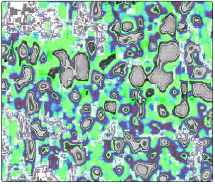
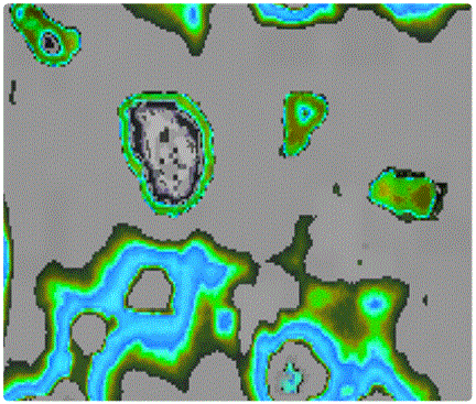
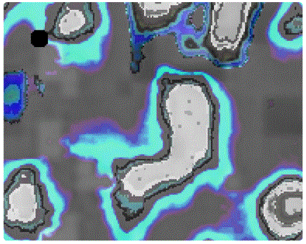
This study refers to what was found as a consequence of processing an image of retinal OCT, and especially with reference to the pigmentary epithelium, through sequencing, desconvolution and filtering of images of the pigment epithelium, adding the adhesion mechanisms and intercelular tension spaces. (Tensioactive intercellular phase).
The morphometric analysis through pixelography, pixelometry and pixelo arquitectura demonstrates in 2D the physical construction of the cells and their intercelular spaces, absolutely original images that are related to similar ones observed in other recent communications that we have made.
With deconvolution, we perform a method operation to restore signals and degraded data by the physical processes of the tomograph, correcting the focuses and noise by diffraction and photons.
The tissue in the study area was cut into small parts of more than 4 to 6 cells, taking independent images and adding the results, achieving it erativea lgorithms such as máximum like hood estimation in microscopy (Guedel).
Introduction
There is a new paradigm in morphological imaginary: it emerges from the intersection of physics, biology and informatics: information as the primary foundation of the universe we perceive and perhaps matter and energy as its representation or unfolding.
It is the fusion of physics with computer theory and genetics. A new understanding of what is the fundamental substrate of the universe: information. Are theoretical informatics in origin, and this is a participatory university. The whole universe is then seen as a computer a cosmic information processing machine. When photons and electrons and other particles interact, what are they actually doing? Inter changing bits, pixels, transmitting quantum states, processing information. The laws of physics are algorithms.
However, the possibility of storing their own information, a unit, which is the pixel, makes it more accurate, dynamic and reliable. Quantum physics is the physics of the possibilities of change, management of unified fields of the four forces: gravity, electromagnetism and the strong and weak force of the atomic nucleus.
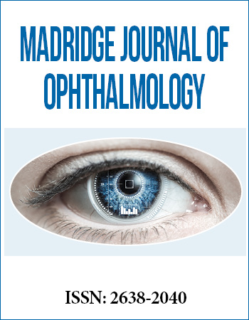
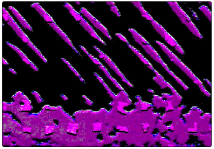
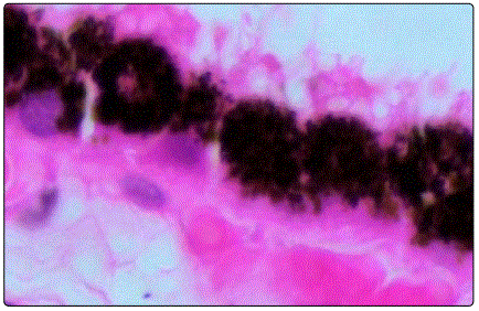
Einstein suggested in his theories, the existence of a field holding space-time transformations and mass - energy. This field is the pixel. In the different photos that we show, can be seen digital sequentiation of the retina tissue, detection technique based in sequencing images, obtained in our Digital Laboratory (Maimonides University).
This image of phisical cell are the definitive methods of physical-mechanical comprehension of the celular activity of the retinal pigment epithelium and the maintenance of the inter phase between cell and cell thereof. On the other hand its function of phagocytosis and its metabolic frame work.
The observed images are produced by digital optical biopsy [1,2], that is, the sequencing of OCT images (optical coherence tomography), to cyto-histology by pixelography, pixelometry and pixelo arquitectural techniques [3-9].
References
Contact us for any additional information - contact@madridge.org
 Madridge Publishers is licensed under a Creative Commons Attribution 4.0 International License.