Research Article
Relationship between Chronic Obstructive Pulmonary Disease (COPD) and C- Reactive Protein (Crp)
Director, Organization Royal Hospital, Oman
*Corresponding author: Nasser Al Busaidi, Director, Department of Medicine, Organization Royal Hospital, Oman, E-mail: enhsa66@yahoo.com
Received: July 2, 2018 Accepted: July 13, 2018 Published: July 19, 2018
Citation: Al Busaidi N. Relationship between Chronic Obstructive Pulmonary Disease (COPD) and C-Reactive Protein (CRP). Madridge J Intern Emerg Med. 2018; 2(2): 71-75. doi: 10.18689/mjiem-1000115
Copyright: © 2018 The Author(s). This work is licensed under a Creative Commons Attribution 4.0 International License, which permits unrestricted use, distribution, and reproduction in any medium, provided the original work is properly cited.
Abstract
Chronic obstructive pulmonary disease (COPD) is a major worldwide health problem which leads to expiratory air-flow limitation which is often slowly progressive over the years. C-Reactive Protein (CRP) usually elevated in patients with COPD and used as a marker for inflammation.
Objectives: The objective of the study to see whether CRP correlated with the severity of COPD in the form of lung function test.
Method: This is a retrospective study, from patients with COPD followed up in a tertiary hospital between Jun 2011 to Dec 1016 who have both CRP measurement and spirometry in same period.
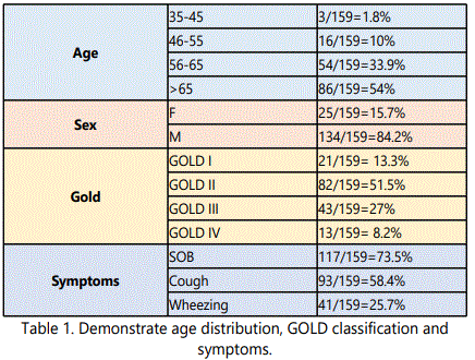
Result: Total number of patient were 159 (84.2% male), mean age was 66 years, most of the patients presented with dyspnea (>73%) followed by cough (>58%) and about quarter of the patient with wheezing. Based on Global Initiative for Obstructive Lung disease(GOLD) classification; most of the patient belong to GOLD II (51.5%), then GOLD III (27%), then GOLD I (13.3) and only 8.2 belong to GOLD IV. Majority of our patients (73%) had raised CRP level (> 5 mg/L), our study showed no significant difference between CRP and GOLD stages but there is a linear negative correlation between FEV1% and CRP level and we found each unit increase of CRP level, there will be decrease of FEV1 by 0.001. This finding is novel as it hasn’t been documented elsewhere.
Conclusion: our study shows that there is a linear negative correlation between FEV1% and CRP level in stable COPD patients.
Keywords: Retrospective study; Spirometry; Obstructive airway disease; Chronic Obstructive Pulmonary Disease (COPD); C-Reactive Protein (CRP); Global Initiative for Obstructive Lung disease.
Introduction
Chronic obstructive pulmonary disease (COPD) is a major health issue worldwide; COPD is a slowly progressive disease over years due to expiratory airflow limitation [1], [2]. The expiratory airflow limitation is generally progressive; though it’s variable from one patient to another; and in most of the cases it is associated with exaggerated inflammatory response of the airways to different particles and gases [1]. The circulating serum level of the inflammatory marker C reactive protein (CRP) are usually elevated in patients with stable COPD however C-reactive protein (CRP) is often used as a clinical marker of acute systemic inflammation such as acute exacerbation of COPD [3]. CRP measurements may provide a prognostic indicator which could give better prediction than the traditional parameters to assess mild to moderate stable COPD, and hence the measurement of CRP may help to detect more accurately the patients at high risk of mortality or morbidity [3].Morrten Dahl and his colleagues found CRP serum level is a strong and independent predictor of future COPD outcomes in patients with chronic obstructive airway disease [4]. Funda Aksu found similar result that CRP correlate well with COPD severity and also he found elevated CRP correlated with COPD and concomitant systemic hypertension and pulmonary hypertension and this association independent of ever-smoking [5]. However, Silva in case control study found serum CRP in COPD has no significant difference when compared with control group [6]. Torres reported in 218 stable moderate to severe COPD patients the level of CRP level was not associated with survival compared with other prognostic clinical tools such as the BODE index, modified Medical Research Council scale, 6-min walk distance and percentage of predicted FEV1 [7]. Zhang in meta-analysis found Patients with COPD had higher serum CRP concentrations than healthy controls in addition, patients with severe COPD had higher serum CRP concentrations than those with moderate COPD but there was no significant difference in serum CRP concentration between patients with mild and moderate COPD [8].
Objective
The objective of our study to assess the level of C Reactive Protein (CRP) and whether it correlate to the severity of COPD among GOLD classes based on spirometry.
Methods
Study population
This is a retrospective study of patients diagnosed with chronic obstructive pulmonary diseases (COPD) based on physician diagnosis at Royal hospital; largest tertiary hospital in Muscat belong to Ministry of Health (MOH), the data was retrieved from Al Shifa (electronic system). The diagnosis of COPD was based on history (symptoms; cough, shortness of breath and or wheezing) and confirmed with spirometry based on GOLD guideline (FEV1/FVC less than 70%) [9]. The patients belong to different GOLD classes (I-IV), we selected patients with diagnosis of COPD and have both investigations done; Spirometry and C- Reactive Protein (CRP) we excluded all patient with Asthma or Asthma and COPD overlap syndrome and patients with Bronchiectasis. After all, 159 patients had been followed up as an out-patients at Royal hospital between period of Jun 2011 and Dec 1016.
The study approved by Royal Hospital ethics committee.
Estimation of CRP
The serum CRP levels was estimated at Royal Hospital laboratory with, the values were considered normal if less than 5 mg/l and abnormal if values were 5mg/L and above, the blood sample obtained when patients has no symptoms suggest acute exacerbation; however there are few patients the blood sample obtained for CRP at the end of recovery of acute exacerbation.
Spirometry evaluation
Spirometry was also done at Royal hospital in the lung function laboratory with ATS specifications, the test was selected close proximity in the timing of CRP measurement as most of the patients had spirometry in different hospital visits, the selected spirometric values coincide with CRP measurement is not postbronchodilator test as in all patient these are a follow up spirometry and not spirometry in the first visit.
Statistical Analysis
Results are presented as number, percent, mean and standard deviation in the form of tables and figures. The continuous data was analyzed by One-Way ANOVA. Relationship between CRP and GOLD stages was analyzed by correlation analysis and linear regression after Log10 transformation.
Result
Total number of patients was 159 out of which 84.2% were male, the mean age + standard deviation 66.2 + 9.7 years, GOLD classification; most of the patient belong to GOLD II (51.5%), followed by GOLD III (27%), followed by GOLD I (13.3) and only 8.2 belong to GOLD IV. Most of the patient presented with shortness of breath (73.5%) followed by cough (58.4%) and about one quarter of the patient presented with wheezing; table 1. The characteristics of female patients are similar to male patients in our population in the form of age, symptoms and also the level of C- reactive protein level, however, this comparison is difficult to make as the number of female patients in our study was less than 16% of the patients.
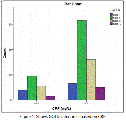
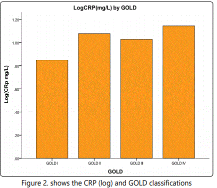
CRP measurement showed as follow; 73% of the patients has CRP(5 mg/l and the rest of the patients <5 mg/l; the median value of CRP was 10.7 mg/l, CRP values based on GOLD classification as follow; GOLD I 62% of patients had CRP≥5 mg/l and rest of the patient their CRP<5mg/l, GOLD II 74.4%of patients had CRP≥5 mg/l and rest of the patient their CRP<5mg/l, GOLD III 74.5% of patients had CRP≥5 mg/l and rest of the patients their CRP<5mg/l and GOLD IV 77% of the patients had CRP≥5 mg/l and rest of the patients their CRP<5mg/l as shown figure 1. Since the distribution of CRP was highly right skewed, therefore a Log10 transformation was applied to make the data normal. Thereafter One-Way ANOVA was done, keeping the GOLD classification as the explanatory variable and the Log10 (CRP mg/L) as the dependent outcome variable. The results yielded an insignificant P-value of 0.226. However, the bar graph (see figure 2) clearly shows that there appears to be a positive correlation between severity of GOLD and CRP mg/L. The reason for the insignificant P-value could be the low sample size of 13 in the GOLD IV category.
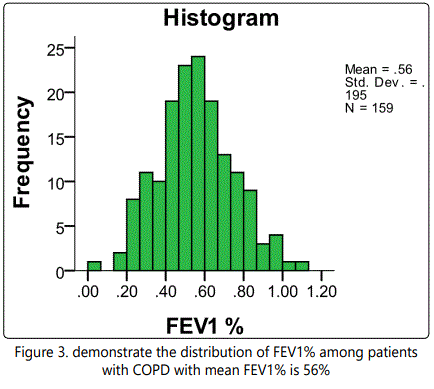
FEV1% was spread as bell shaped distribution as shown in figure 3 with mean ± standard deviation= 56 ± 19.5%. A correlation analysis was conducted to examine the correlation between FEV1% and CRP level, the FEV1% being the dependent variable. The correlation coefficient was found to be -0.162, which shows a weak negative correlation. However, the linear model was found to be statistically significant, F (1,157) = 4.224, P-value= 0.042. The linear regression equation turns out to be: FEV1% = 0.577- 0.001 x CRP. The model predicts that that for a unit increase in the CRP level, there will be decrease of FEV1 by 0.001.
Discussion
In this retrospective study of 159 patients with COPD followed up in tertiary hospital between June 2011 and December 2016, more than 84% of the patient are male, this male to female distribution in our population is different from other population as reported by Pinto-Plata from Poston in United State of America as in his COPD patients was 59% male 10 and 55% male as reported by Morten Dahl and his colleagues in European populations 4, these differences in male to female ratio is expected in our society as women are much less smoker then women in western countries. The mean age 66 years and that is same age as reported by PintoPlata [10]. In United State of America and same as reported by Morten 4 in European populations. C-Reactive Protein (CRP) was high (>5) in more than three quarter of COPD patients, these fact is well reported in many studies, Zhang analyses 17 studies in a meta-analysis, all studies except one by Kersul et al 2011, showed significant association between CRP and COPD 8 this association thought to be due to bacterial colonization in stable COPD patients [11]. Takemura reported that COPD patients whom CRP high might have Chlamydia or Mycoplasma infection [12]. However, Anderson thought that elevated CRP in stable COPD patients unlikely due to smoldering chest infection [13].
In our study population all patients presented with either shortness of breath, cough and or wheezing. Shortness of breath was 73% followed by cough in 58% of the patient and in about one quarter has wheezing. Majority of the patients had combinations of these symptoms, these finding are compatible with other studies [14-16]. Gallego reported there is weak relationship between intensity of dyspnea assessed by Medical Research Council (MRC) scale and predicted FEV1 [17]. Smith reported the cough is very common symptom among patients with COPD and the frequency of cough vary according to the age group ranging from less than 20% in younger age (35-40 years) up to 50% in the age group above 60 years, she also reported that cough correlated poorly to the severity of airway obstruction [18]. In a telephone survey of 2950 COPD patients Kessler et al. found that cough was reported by 55% of subjects with 20% rating it severe to extreme [19].
73% of our patients have their CRP higher than normal (≥ 5mg/l), GOLD classification in our population has little impact in the level of CRP; GOLD I, II, III and IV, 62%, 74.4%, 74.5% and 77% respectively, the results yielded an insignificant P-value of 0.226, however, the bar graph (see figure 3) clearly shows that there appears to be a positive correlation between severity of GOLD and CRP mg/L. The reason for the insignificant P-value could be the very low sample size of 13 in the GOLD IV category, this weak correlation between GOLD and CRP was also reported by other researchers as it was reported no statistically significant difference between serum concentrations in mild and moderate COPD (WMD = 0.67 mg/l, 95% CI –0.08, 1.42, P = 0.08 [20-23]. As it was clearly reported by Y Zhang in his meta-analysis [8]. However Nena reported. The level of CRP showed significant correlation with the severity of clinical presentation according to GOLD classification. The higher the CRP concentration, the higher was the disease severity determined according to GOLD classification (p<0.001) [24], Simonovska also reported significant statistically difference between mean values of CRP and GOLD categories [25]. Similar result also reported by Nillawar in group of patients from India and the result was statistically difference between mean value of CRP and GOLD IV when compared with GOLD II or GOLD III with P = 0.0089 and P = 0.03 respectively [26]. However Halvani from Iran showed similar to our result that the mean CRP level in COPD patient was not significantly correlated with severity of disease [27].
The correlation between FEV1% and CRP level, the FEV1% being the dependent variable, the correlation coefficient was found to be –0.162, which shows a weak negative correlation. However, the linear model was found to be statistically significant, F (1,157) = 4.224, P-value= 0.042. This negative correlation between FEV1% and CRP was reported by searchers. Aksu found CRP levels significantly correlated with age, FEV1% predicted, FVC% predicted, SpO2 , MMRC, 6-minute walk distance, BODE scores and hemoglobin levels [5]. Similar result also reported by Shrivastava as he demonstrated clearly that C-reactive protein levels correlate well with FEV1% (p0.029) and GOLD stage of disease (p0.040) [28]. This negative correlation between lung function test parameters and CRP level was reported by many other authors [11], [29-32]. However, Abolhassan Halvani reported there is no statistically significant negative correlation between CRP and severity of COPD assessed by lung function test in 45 COPD patients [27]. Halvani hypothesize this result due the use of Inhaled corticosteroid uses in this group of patients.
In our study we concluded that the linear regression equation turns out to be: FEV1% = 0.577- 0.001 x CRP, this model predicts that that for a unit increase in the CRP level, there will be decrease of FEV1 by 0.001.
This novel finding, haven’t been mentioned by any researchers before, however, being the study population 159 patient we need a larger number of patients to confirm this finding.
Tables & Figures


Conclusion
In this study is retrospective study which includes 159 stable COPD patients with average age 66 years’ old were are majority are male (84.2%), shortness of breath is the most common symptoms (73.5%) GOLD II was most dominant (51.5%) followed by GOLD III (27%) and only 8.2% of our patients belong to GOLD IV, majority of our patient (73%) had raised CRP (≥ 5 mg/l), however, in our study showed no significant different between CRP and GOLD stages but there is linear negative correlation between FEV1% and CRP level and we found each unit increase of CRP level, there will be decrease of FEV1 by 0.001. This finding is novel as it hasn’t been found in the previous studies.
Declaration of Interest: None of the authors declared any conflict of interest regarding this study.
References