Case Report Article
Myxofibrosarcoma of Nasal Cavity and Sinuses
Department of Head and Neck Surgery, the Second Affiliated Hospital of Nanchang University, Nanchang 330006, Jiangxi, China
*Corresponding author: Hong-Bing Liu, Department of Head and Neck Surgery, The Second Affiliated Hospital, Nanchang University, 1# Minde Road, Nanchang Jiangxi, China, Tel: +86-0791-86295805, Fax: +86-0791-86262262, E-mail: liuhb1992@163.com
Received: June 25, 2018 Accepted: June 29, 2018 Published: July 5, 2018
Citation: Zhang JX, Liu HB, Huang GJ. Myxofibrosarcoma of Nasal Cavity and Sinuses. Madridge J Immunol. 2018; 2(1): 49-50. doi: 10.18689/mjim-1000111
Copyright: © 2018 The Author(s). This work is licensed under a Creative Commons Attribution 4.0 International License, which permits unrestricted use, distribution, and reproduction in any medium, provided the original work is properly cited.
Abstract
Myxofibrosarcoma (MFS) is a low degree of fibrous tissue malignant lesions. It usually occurs in the limbs but extremely rare in the nasal cavity and sinuses. We herein present a case of nasal MFS. A 41-year-old male patient presented with a chief complaint of a right stuffy nose and shed tears for almost two months. Since the tumor has high recurrence rate, mainly infiltrative growth, locally extended resection is the basic treatment. Postoperative adjuvant radiotherapy and chemotherapy can reduce the recurrence rate and metastasis rate.
Keywords: Nasal cavity; Sinuses; Myxofibrosarcoma.
Introduction
Myxofibrosarcoma previously known as myxoid malignant fibrous histiocytoma, is considered one of the most common fibroblastic sarcomas of the elderly, predominantly affecting patients between fifty and eighty years of age and mainly in the limbs. We report the case of nasal myxofibrosarcoma of 41 years old, surgically resected.
Case Report
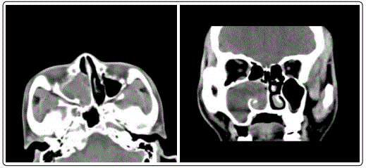
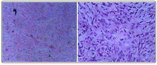
An 41 years old man with right nasal congestion for two months. Began to appear without apparent inducement right nasal obstruction patients nearly 2 months ago, was persistent, progressive increase, accompanied by headache and nasal, smell decline, no fever, nasal bleeding facial numbness and pain. Physical examination: nasal septum slightly left, right nasal see large amounts of red light with smooth surface and new biological purulent secretions. Before admission to hospital in nasal biopsy, pathological examination showed the right nasal fibroma. The relevant examination after admission, sinus CT (Figures 1 and 2) in soft tissue. The right maxillary sinus and the nasal cavity filled nasal soft tissue protrusion backward nostril, maxillary sinus enlargement, sinus wall bone damage. Under general anesthesia by nasal endoscopic resection of the right nasal cavity tumor and the anterior lacrimal fossa in right maxillary sinus tumor, Postoperative pathology (Figures 3 and 4) see yellow gray red tumor tissue, which arc shaped vascular tumor cells showed different degrees of atypia. The diagnosis was consider myxofibrosarcoma. Immunohistochemistry showed that the tumor cells of CK(±), Vim(+), BCL-2(+), S100(-), SMA(-), CD34(-), ALK(-), Des(-), CD99(-) Ki-67 about 30%(+). Postoperative given anti-inflammatory and hemostasis treatment, postoperative 4th day oncology follow-up treatment.


Discussion
Myxofibrosarcoma is a kind of malignant tumor of fibrous tissue, the lesions occur in limbs in nasal cavity and paranasal sinuses are extremely rare. Myxofibrosarcoma accounted for about 10% of the meat is soft tissue, malignant tumor derived from fibrous tissue [1], the main manifestations of the slow growth of the local painless mass [2]. The 2002 edition of the WHO classification of the MFS are defined to include a series of malignant fibrous mother cell lesions, not the same amount of myxoid matrix, cellular pleomorphism and unique curved blood vessels, and which included myxoid [3].
Myxofibrosarcoma usually occurring in 50-80 years old people. Mainly occurs in the limbs, lower limbs than upper limbs, followed by the trunk [4]. This report is in nasal cavity and sinuses myxofibrosarcoma, and the patient is middle-aged male, 41 years old, rarely reported in the literature of the tumor in the nasal cavity and sinuse. The study found that the tumor was mainly composed of spindle shaped cells arranged in fascicles which showed nodular growth and infiltration of surrounding tissue, tumor cell atypia and polymorphism performance, visible from the low level to a high level of continuous tumor cell lineage, cell sparse zone mucilage matrix curve showed the typical vascular stroma [5]. Immunohistochemistry: vimentin is positive, cells markers (CD68, Mac387, FXba) and S-100 protein are negative. The characteristics of myxofibrosarcoma clinical manifestation and imaging lack of specificity, mainly depends on the pathological diagnosis. Myxofibrosarcoma can be seen in uniform spindle the shape of cell image and herring bone like cells showed a general beam, immune markers is negative. Nerve fibroma often have a myxoid stroma, and a bundle of collagen bundles. MFS morphological characteristics are as follows: diffuse myxoid interstitial, surrounded by fibrous septa incomplete; tumor cells are rare, loosely arranged, showed short fusiform shape, star, can also be pleomorphic or foam. The rich slender arc-shaped vascular network. Tissue pathology has the different manifestations of the most high differentiation of polymorphic sarcoma from the low differentiated mucinous sarcoma of the cell [3].
Reported in the literature about 50% - 60% of the patients had local recurrence of myxofibrosarcoma, about 20% - 25% appears distant metastases [6]. Risk factors that lead to poor prognosis include tumor necrosis, tumor diameter >5 cm and the reduction of mucus area. Because of the high recurrence rate, mainly infiltrative growth, the extended local excision is the basic treatment. Adjuvant chemotherapy can reduce the recurrence rate and metastasis rate after operation. The diagnosis rate of the disease and the survival rate of the patients were improved after surgery and radiotherapy and chemotherapy. The postoperative recurrence and metastasis were also found in the early postoperative period. Because the tumor recurrence rate is high, mainly in invasive growth, it is the basic treatment of local expansion.
References
- Ferrari A, Sultan I, Huang TT. Soft tissue sarcoma across the age spectrum: a population-based study from the Surveillance Epidemiology and End Results database. Pediatr Blood Cancer. 2011; 57(6): 943-949. doi: 10.1002/pbc.23252
- AngerVall L, Kindblom LG, Merck C. Myxofibrosarcoma. A study of 30 cases. Acta Pathol Microbiol Scand A. 1977; 85(2): 127-140. doi: 10.1111/j.1699-0463.1977.tb00410.x
- Mentzel T, Calonje E, Wadden C. Myxofibrosarcoma. Clinicopathologic analysis of 75 cases with emphasis on the low-grade Variant. Am J Surg Pathol. 1996; 20(4): 391-405. doi: 10.1097/00000478-199604000-00001
- Nascimento AF, Bertoni F, Fletcher CD. Epithelioid variant of myxofibrosarcoma: expanding the clinicomorphologic spectrum of myxofibrosarcoma in a series of 17 cases. Am J Surg Pathol. 2007; 31(1): 99-105. doi: 10.1097/01.pas.0000213379.94547.e7


