Research Article
Radiographic Evaluation of Third Mandibular Molar Development as an Age Indicator in a Swedish Population
Department of Dental Medicine, Division of Orofacial Diagnostics and Surgery – Image and Functional Odontology, Huddinge, Karolinska Institute, Stockholm, Sweden
*Corresponding author: Liljana Simonsson, Department of Dental Medicine, Division of Orofacial Diagnostics and Surgery-Image and Functional Odontology, Huddinge, Karolinska Institute, Stockholm, Sweden, E-mail: liljana.simonsson@bredband.net
Received: August 19, 2016 Accepted: September 13, 2016 Published: February 16, 2017
Citation: Simonsson L, Näsström K, Kullman L. Radiographic Evaluation of Third Mandibular Molar Development as an Age Indicator in a Swedish Population. Madridge J Dent Oral Surg. 2017; 2(1): 31-37. doi: 10.18689/mjdl-1000108
Copyright: © 2017 The Author(s). This work is licensed under a Creative Commons Attribution 4.0 International License, which permits unrestricted use, distribution, and reproduction in any medium, provided the original work is properly cited.
Abstract
The refugee situation in Sweden today has been the most difficult since Second World War. For unaccompanied children without any identity documentation it is important to show that they are below 18 years old, giving them a chance to get asylum.
The purpose of the study was to evaluate the development of the third molar in a sample of Swedish population.
A total sample of 1031 panoramic radiographs was analyzed. The subjects were 12-25 years old. The mineralization stages of the third mandibular molar were assessed according to Demirjianʼs method. Age ranges were wide for the different developmental stages. By the age of 20, most of the teeth were fully developed.
In conclusion, the third mandibular molar is the only usable tooth for age estimation around the age of 18 years, although it is the most variable tooth in the dentition. The data described may provide reference for age assessment.
Keywords: Radiographic Evaluation; Mandibular; Molar; Panoramic Radiographs.
Introduction
Age estimation in humans has taken an important role in todayʼs modern society [1-4]. It is of great importance in several areas: deciding the age of living persons and in the identification process of unknown deceased persons. For unknown deceased persons, it is of significance to identify the victims of mass disasters, fires, accidents, crashes and homicides. As regards the living persons, it is of significance when the birth certificate is not available. Age estimation includes requirements in assessing whether a person has reached the age of criminal responsibility in cases like rape, kidnapping, employment, marriage, adoption and illegal immigration.
Today there is an increasing number of immigrants in western countries due to economic globalization, wars, conflicts and disasters in different countries [5]. Many of them are without valid identification papers. The authorities in many countries may often be in doubt regarding their uncertain age due to legal consequences [1,2,4,6]. One of the reasons is that children under the age of 18 have special rights and benefits according to the UN Child Convention and cannot be sent back to their countries of origin [7], which results in an increased chance for them of being granted asylum. Other reasons might be civil or criminal proceedings.
In Sweden, some of the ages of legal relevance are 15 and 18. Adolescents between 15-18 years of age are considered juvenile delinquent in the state of the law and will face a milder punishment in cases of criminal act. By the age of 18 one has reached the age of criminal responsibility [8].
There are some recommendations in forensic age assessment proposed by the international and interdisciplinary Study Group on Forensic Age Diagnostics (AGFAD) [9]. They include a) a physical examination, including body measurements and assessments of signs of sexual maturity, b) X-ray examination of the left hand and c) a dental evaluation, including clinical examination of the dental status and X-ray examination. If the skeletal development of the hand is completed, an X-ray examination of the clavicles needs to be performed additionally.
In Sweden by tradition, the age estimation has been channeled by The Swedish Migration Board [10]. The recommendations regarding age assessment for children and juveniles seeking asylum have been set by the National Board of Health and Welfare [11]. The age assessment was commenced by clinical examination of a pediatrician. In cases where additional information was needed, could an additional x-ray of the hand and x-ray of the teeth be requested. However, the refugee situation in Sweden today has been the most difficult since world Second World War [12-15]. Sweden has taken far more refugees per capita than any country in EU. Some of the reasons are the war in Syria and the turbulence in Middle East and Africa. During 2014, more than 80 000 came to Sweden seeking asylum. In 2015, the number had more than doubled. During the first three months in the year 2016, almost 10 000 refugees had fled to Sweden seeking asylum, which is more than 1000 refugees per week. Out of these, were more than 10% unaccompanied children. If an unaccompanied child doesnʼt have any identity documentation and if there is a doubt regarding the individualʼs age has the Migration Board to decide whether the person is under or over the age of 18. A child which is below 18 has the right for being granted asylum. Today, has the applicant the burden himself to proof his or her chronological age and the methods available to him or her is identification documents, his or her statement and/or a medical examination including dental and skeletal radiograms. This examination, which involves dental and wrist-bone x-rays, can usually help in narrowing the age span of (young) adolescents.
While the skeletal and somatic developments are influenced by the environment, nutrition, hormonal imbalance and diseases, teeth seem to be less affected [16-19]. Teeth are also highly resistant to severe insults such as chemicals, fire, cold and heat [20-22]. While several age estimation modalities are invasive, requiring lengthy processing times, use of expensive sophisticated equipment or services of an experienced pathologist when using histological methods [23-26], radiological evaluation of the development of the teeth is a simple, quick and non-invasive investigation method used in-vivo. Teeth are useful predictors, especially during early years when many teeth are developing, up until the age of 14-15 years [27-31]. Third molars are less used for dental age determination due to their variability in time of formation and time of eruption [32]. However, beyond the age of 14-15, the only teeth still in development are third molars [29,33-35]. That is the reason why radiographic assessment of third molar formation is an important method in forensic sciences.
A number of studies, of which all have used Demirjianʼs method, have shown a population-specific third molar development among different geographical groups: Korea [36], Iran [37], Portugal [38], Spain [39], India [40], UK [41], Canada [42], western China [43], USA [44], Turkey [45], Brazil [46], Germany [47], Thailand [48], Malaysia [49]. The above mentioned studies have revealed slight differences in mineralization stages of third molars between different populations. Very few studies have been implemented among Swedish juveniles [30,34,50]. Hence, it was considered of importance to determine thirdmolar developmental stages in a Swedish sample population to create a reference for chronological age estimation.
Material and Methods
Exclusion criteria were (i) patients with known systemic or developmental disease, (ii) panoramic radiographs of patients without any of third mandibular molar present, (iii) radiographs with distortions in the selected region/unclear panoramic radiographs and (iiii) pathology in the third mandibular area in the radiographs (for example cysts).
The study design was a retrospective cross-sectional study. A total of 1031 panoramic radiographs from patients of ages between 12 and 25 years were analyzed. The ethnicity of the study population was not recorded, since there are several mixed marriages and adopted children with Swedish family names. The radiographs were collected from Karolinska Institute, Division of Image and Functional Odontology, Department of Dental Medicine, Huddinge), Stockholm, from patients attending the school between 2011–2012. All radiographs had previously been taken for a diagnostic and treatment purpose and were digital. A Planmeca Promax 2D panoramic machine (Helsinki, Finland) was used. The radiographs were stored and analyzed using Romexis version 2.2.2R software.
The radiographs were coded during assessment, so that the dates of birth and exposure were unknown to the examiner. Patient identification number, sex, date of birth, date of X-ray and mineralization stages of the lower third molars had been earlier recorded. Each patientʼs age was calculated as the date of X-ray minus the date of birth and recorded as years and parts of years. Microsoft Access tables were used for the registration of data. The developmental dental stage in relation to the age and gender specific differences was studied.
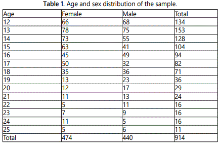
The age and sex distribution of the study population is shown in table 1.
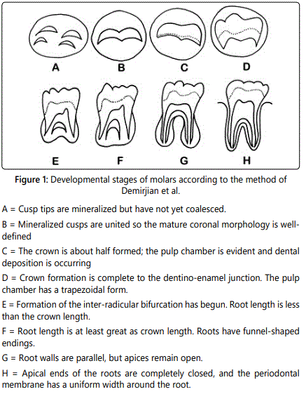
The mineralization of the third mandibular molar was assessed according to the method described by Demirjian et al [27] consisting of 8 stages as described in figure 1.
The study was restricted to the evaluation of the lower third molars because the assessment of the third molar of the upper jaw may be problematic as the wisdom tooth is often superimposed over other anatomic structures. It has also been found that, the reproducibility (intra observer reliability) is higher for mandibular third molars than maxillary third molar [51].
Assessment of the radiographs included in the study was performed independently by one inexperienced examiner (L.S.) without any knowledge of the subjectʼs chronological age and gender. However prior to the study, a training sample of panoramic radiographs was assessed with an experienced examiner (L.K). Inter-observer reproducibility was assessed on 46 independent samples of radiographs, expressed as Cohenʼs kappa coefficient. Intra-observer reliability was tested by reexamining 90 radiographs (10% of the radiographs) by the same observer a month after the initial examination (L.S.).
Statistical analysis was performed using SPSS 21.0 (SPSS Inc, Chicago IL) for Windows. The data regarding differences between 38 and 48 was analyzed using Spearman correlation coefficient and t-test for pair wise comparison. Statistical data including maximum, minimum and mean age as well as standard deviation of the ages were obtained for each stage of mineralization of both sexes. For each mineralization stage the difference in mean age between the sexes was analyzes separately with independent t-test. The results were considered statistically significant when the p-value was < 0.05.
The ethical approval was received by the Regional Ethic Board in Stockholm, Sweden, and performed in accordance with the Helsinki declaration, registration number 2013/906/31.
Results
Inter-observer agreement was excellent: correlation coefficient (r) was 0,99. Very good agreement was also indicated by the intra-observer result, correlation coefficient (r) was 0,91.
The initial sample consisted of 1031 patients. Out of these patients, 113 had none of their lower third molars (12.4%), two had pathology on both sides, in one panoramic radiograph was distortion found on both of the third mandibular molars and in one panoramic radiograph were extraction alveoli present on both sides in the third molar region. 53 of all the included patients had only one mandibular molar (5.8%). After removal of the radiographs based on the exclusion criteria, a total sample of 914 patients (440 male and 474 females) was included in the study.
Comparisons between the left and the right side of the third mandibular molars did not show any statistically significant difference. Due to this observation, we pooled the results for both sides, 38 and 48, for each gender in our tables (tables 2-4).
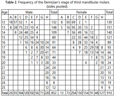
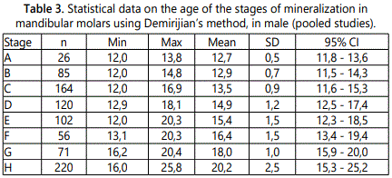


Table 2 show the distribution and the frequency of different mineralization stages according to Demirjian, separately for each tooth for both males and females. In this sample, at a certain age the difference in the maturation of the wisdom tooth could be as much as 6 mineralization stages. The minimum age for apex closure (stage H) is 16 in males, respectively 17 in females. Beyond the age of 20, all of the third mandibular molars have finished their root formation (table 2).
Tables 3 and 4 shows the result for each tooth mineralization stage according to the specific ages, presented separately for males and females. Results are expressed as minimal and maximal age, mean values, standard deviation (SD) and ± 2 SD for girls and boys respectively. For individuals 18 years and older, it was seen that they could be in all the mineralization stages between D-H for both males and females.
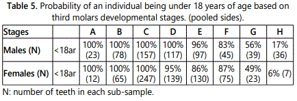
Table 5 displays the probability of an individual being less than 18 years old based on third molar development stages. In every stage, except for stage F, the probability was higher for the males of being younger than 18 years old, compared to the females, based on each sub-sample in the specific development stage.
Discussion
In past, there have been several methods to assess dental development [27-30,33,50,52-56]. The advantage of using few tooth development stages in age estimation is better reliability of inter-examiner agreement, but this results in less accuracy. Demirjianʼs 8 stage model is widely used for its simplicity and objectivity [38,40,42-45]. This method also shows very good intra- and inter-examiner agreement [45,51,57] and the methodical errors are small [58]. It does not rely upon measurement length on radiographs, which sometimes can be distorted, but rather on development stages of the tooth: by estimating the form of the tooth bud and the relation of crown/root length. Out of several tested methods it has been found that Demirjianʼs method is the most accurate in terms of evaluation of mineralization of the third molar [55,57].
The bilateral absence of third mandibular molars in our study is slightly above the one found by Levesque [32]: 12.4% compared to up to 9% in their study.
Left-right asymmetry in the tooth formation is common for the third molar according to Nolla, more than elsewhere in the dentition [52]. However, no statistical differences were found in our study between mineralization of right and left third molars. This finding is consistent with other studies as well [37,59-61].
Regarding differences in gender, the pattern of development for the first seven forming teeth is that they form earlier in females than in males [27,33]. The reverse is seen to occur in the third molar formation and eruption, which is shown in several studies [33,37,38,42]. The present work has shown statistically significant differences in the mean age between males and females mainly in stages C, D and E. The males reached these stages earlier than females. The highest difference was seen at stage E in our study, where males were 9 months ahead of females. These results are mainly in accordance with other findings [49,50,62,63].
In general, the individual variation in dental development is larger for older ages than in younger children, as found in other studies [27]. Li et al found that individual subjects could have reached up to six different mineralization stages of development, according to Demirjian, in one and the same age group [43]. The same results were seen in our study as well (Tables 2 and 3). In the study of Johan et al, the span was no more than five stages [49].
As seen in table 2, two extremes can be observed in our study. If a subject is graded A to D according to Demirjianʼs 8 mineralization stages, there is little likelihood that the individual is above the age of 18, seen in other studies as well [60,63]. On the other hand, in our study the minimum age for apex closure (stage H) was 16 in males, respectively 17 in females. This means that if a subject is graded H, one can presume that the subject could be at least 16 years of age or more in males respectively 17 in females.
According to the results of the present study, the SD was low in the youngest ages and increased with the higher age groups. These findings however should be interpreted cautiously, because of the cross-sectional nature of the study. If our sample groups would have included younger ages such as 5-6 year old children, when the formation of the crypt of third mandibular molar is seen in the earliest individuals [35], the mean age and SD up to stage E probably would have been affected.
The results of this study have shown that the standard deviation can be up to 1, 6 years, as seen in Table 3 and 4 (if stage H is excluded). This means that the estimated “dental age” can be within an interval of about 3, 2 years, based on 95% of the population in the sample (±2 standard deviations). This data based on the teeth alone is not an optimal solution for age estimation, but has been proven applicable in other studies [34,63]. The third molar is the only tooth that has not yet been mineralized cases when the individual is above the age of 14.
Previous Swedish studies concerning the third mandibular molar mineralization [34,50] were limited in particular by the low number of subjects in the study. Another aspect is the choice of method for assessing the development of the tooth. Most studies in other parts of the world assess the mineralization of the third molar in accordance to Demirjianʼs 8 stages [40,42-44] To the extent that comparisons can be made, Kullman et al [50] found that crown development was ended and root formation initiated at a mean age of 15,0 years in males and 15,4 years in females. The nearest corresponding stage in Demirjianʼs scale is stage D, which in our study occurred at 14,9 years in males respectively 15,3 years in girls. This is very close to the result as in Kullmanʼs study. Stage F by Demirjian is recorded in another Swedish sample in Thorson et al [34], who found the mean age for males to be 16, 6 and 17, 0 in females. That could be compared to 16, 4 in males and 16, 6 in females in our study. The nearest corresponding stage in Kullmanʼs study would be stage 3 or 4, when at least 1/2 or more of the estimated root length has been formed. This occurs at 16, 9 years in males and 16, 8 years in females in stage 3. The stage when the apex closure has been initiated but not completed occurs at 19, 2 years in males and 19, 9 in females in Kullmanʼs study. That could be in comparison with stage G by Demirjian, which in our study occurs at 18, 0 years in both genders, compared to 18, 4 in males and 18, 7 in females in Thorson et al.
Differences found between this sample population and populations in other studies can be attributed to many variables: age structure of the sample, sample size, statistical approach, biological variation of individual children and experience in age assessment of the observer. Nevertheless, the differences in developmental stages of the third molars in various populations call for more ethnic-specified reports to be performed throughout the world. The reason is to get an accurate view of the association between chronological age of the individuals and the developmental stage of the third molars.
There are several considerable advantages in this study. Concerning the different genders and different sizes of the mandibular molar area, the subjects were relatively evenly distributed. Also the number of the subjects included in this study was higher than in other previous Swedish studies.
The present study has three main limitations. First, the subjects in this study were all based on patients from one limited area in Sweden, Stockholm, instead of being selected at random throughout the country. Thus, theoretically, differences between our results and other studies could partly be due to the narrow geographic location. Secondly, only filed radiographs were used in the study, since additional exposure to patients for this particular purpose could not be justified. The random selection of the subjects was therefore limited since only patients referred to Image and functional odontology department in Karolinska Institute were included. There is however no indication that the sample referred to the Karolinska Institute should in differ in a particular way or for some reason in the development of the third mandibular molar, compared to the rest of the population in Stockholm. Third, the ethnicity was not controlled in this study. However, a various population with different ethnical groups in Sweden is partly to be expected. Most of the variations of different groups are expected in the big cities and the southern parts of Sweden.
Our results should be considered with caution. Further studies in different areas in Sweden, also involving the recording of ethnicity, need to be performed in order to confirm the results in this study. Also, larger samples are recommended.
In summary, the radiological development of the third molar may be a useful biological variable for estimating the age of a person between his teens and early 20s. Even though this tooth shows substantial variation in its formation, it could be of high value in the absence of better information. However, it is important to remember that it is recommended by some researchers to combine the data from the wisdom tooth mineralization with other assessed medical examination findings, such factors as skeletal maturity, in order to minimize the margin of error for the age estimation. Combining of important factors, such as using multiple regression methods, might reduce the range of variation of the overall estimation to a certain extent.
Conflicts of Interest: The authors declare no conflicts of interest with this submission.
References
- Solheim T, Vonen A. Dental age estimation, quality assurance and age estimation of asylum seekers in Norway. Forensic Sci Int. 2006; 159: S56-60. doi: 10.1016/j.forsciint.2006.02.016
- Schmeling A, Olze A, Reisinger W, Rösing FW, Geserick G. Forensic age diagnostics of living individuals in criminal proceedings. Homo. 2003; 54(2): 162-9.
- Ritz TS, Cattaneo C, Collins MJ, Waite ER, Schütz HW, Kaatsch HJ, et al. Age estimation: the state of the art in relation to the specific demands of forensic practise. Int J Legal Med. 2000; 113(3): 129-36.
- Schmeling A, Reisinger W, Geserick G, Olze A. Age estimation of unaccompanied minors. Part I. General considerations. Forensic Sci Int. 2006; 159: S61-4. doi: 10.1016/j.forsciint.2006.02.017
- United Nations High Commissioner for Refugees (UNHCR). Asylum Levels and Trends in Industrialized Countries. 2006.
- Schmeling A, Olze A, Reisinger W, Geserick G. Age estimation of living people undergoing criminal proceedings. Lancet. 2001; 358(9276): 89-90. doi: 10.1016/S0140-6736(01)05379-X
- Schmeling A, Grundmann C, Fuhrmann A, Kaatsch HJ, Knell B, Ramsthaler F, et al. Criteria for age estimation in living individuals. Int J Legal Med. 2008; 122(6): 457-60. doi: 10.1007/s00414-008-0254-2
- Gron AM. Prediction of tooth emergence. J Dent Res. 1962; 41: 573-85. doi: 10.1177/00220345620410030801
- Cameriere R, Flores MC, Mauricio F, Ferrante L. Effects of nutrition on timing of mineralization in teeth in a Peruvian sample by the Cameriere and Demirjian methods. AnnHum Biol. 2007; 34(5): 547-56. doi: 10.1080/03014460701556296
- Garn SM, Lewis AB, Blizzard RM. Endocrine factors in dental development. J Dent Res. 1965; 44: 243-58.
- Melsen B, Wenzel A, Miletic T, Andreasen J, Vagn HPL, Terp S. Dental and skeletal maturity in adoptive children: assessments at arrival and after one year in the admitting country. Ann Hum Biol. 1986; 13(2): 153-9.
- Patidar KA, Parwani R, Wanjari S. Effects of high temperature on different restorations in forensic identification: Dental samples and mandible. J Forensic Dent Sci. 2010; 2(1): 37-43. doi: 10.4103/0974-2948.71056
- Pramod JB, Marya A, Sharma V. Role of forensic odontologist in post mortem person identification. Dent Res J (Isfahan). 2012; 9(5): 522-30.
- Crubézy E, Amory S, Keyser C, et al. Human evolution in Siberia: from frozen bodies to ancient DNA. BMC Evol Biol. 2010; 10: 25. doi: 10.1186/1471-2148-10-25
- Willems G, Moulin RC, Solheim T. Non-destructive dental-age calculation methods in adults: intra- and inter-observer effects. Forensic Sci Int. 2002; 126(3): 221-6.
- Soomer H, Ranta H, Lincoln MJ, Penttilä A, Leibur E. Reliability and validity of eight dental age estimation methods for adults. J Forensic Sci. 2003; 48(1): 149-52.
- Vandevoort FM, Bergmans L, Van Cleynenbreugel J, et al. Age calculation using X-ray microfocus computed tomographical scanning of teeth: a pilot study. J Forensic Sci. 2004; 49(4): 787-90.
- Fu SJ, Fan CC, Song HW, Wei FQ. Age estimation using a modified HPLC determination of ratio of aspartic acid in dentin. Forensic Sci Int. 1995; 73(1): 35-40.
- Demirjian A, Goldstein H, Tanner JM. A new system of dental age assessment. Hum Biol. 1973; 45(2): 211-27.
- Hägg U, Matsson L. Dental maturity as an indicator of chronological age: the accuracy and precision of three methods. Eur J Orthod. 1985; 7(1): 25-34.
- Moorrees CF, Fanning EA, Hunt EE. Age variation of formation stages for ten permanent teeth. J Dent Res. 1963; 42: 1490-502. doi: 10.1177/00220345630420062701
- Kullman L. Accuracy of two dental and one skeletal age estimation method in Swedish adolescents. Forensic Sci Int. 1995; 75(2-3): 225-36. doi: 10.1016/0379-0738(95)01792-5
- Haavikko K. Tooth formation age estimated on a few selected teeth. A simple method for clinical use. Proc Finn Dent Soc. 1974; 70(1): 15-9.
- Levesque GY, Demirijian A, Tanguay R. Sexual dimorphism in the development, emergence, and agenesis of the mandibular third molar. J Dent Res. 1981; 60(10): 1735-41.
- Haavikko K. The formation and the alveolar and clinical eruption of the permanent teeth. An orthopantomographic study. Suom Hammaslaak Toim. 1970; 66(3): 103-70.
- Thorson J, Hägg U. The accuracy and precision of the third mandibular molar as an indicator of chronological age. Swed Dent J. 1991; 15(1): 15-22.
- Liversidge HM. Timing of human mandibular third molar formation. Ann Hum Biol. 2008; 35(3): 294-321. doi: 10.1080/03014460801971445
- Lee SS, Byun YS, Park MJ, Choi JH, Yoon CL, Shin KJ. The chronology of second and third molar development in Koreans and its application to forensic age estimation. Int J Legal Med. 2010; 124(6): 659-65. doi: 10.1007/s00414-010-0513-x
- Rai B, Kaur J, Jafarzadeh H. Dental age estimation from the developmental stage of the third molars in Iranian population. J Forensic Leg Med. 2010; 17(6): 309-11. doi: http://dx.doi.org/10.1016/j.jflm.2010.04.010
- Caldas IM, Júlio P, Simões RJ, Matos E, Afonso A, Magalhães T. Chronological age estimation based on third molar development in a Portuguese population. Int J Legal Med. 2011; 125(2): 235-43. doi: 10.1007/s00414-010-0531-8
- Prieto JL, Barberia E, Ortega R, Magana C. Evaluation of chronological age based on third molar development in the Spanish population. Int J Legal Med. 2005; 119(6): 349-54. doi: 10.1007/s00414-005-0530-3
- Acharya AB. Age estimation in Indians using Demirjianʼs 8-teeth method. J Forensic Sci. 2011; 56(1): 124-7. doi: 10.1111/j.1556-4029.2010.01566.x
- Boonpitaksathit T, Hunt N, Roberts GJ, Petrie A, Lucas VS. Dental age assessment of adolescents and emerging adults in United Kingdom Caucasians using censored data for stage H of third molar roots. Eur J Orthod. 2011; 33(5): 503-8. doi: 10.1093/ejo/cjq101
- Olze A, Pynn BR, Kraul V, et al. Studies on the chronology of third molar mineralization in First Nations people of Canada. Int J Legal Med. 2010; 124(5): 433-7. doi: 10.1007/s00414-010-0483-z
- Li G, Ren J, Zhao S, et al. Dental age estimation from the developmental stage of the third molars in western Chinese population. Forensic Sci Int. 2012; 219(1-3): 158-64. doi: 10.1016/j.forsciint.2011.12.015
- Lewis JM, Senn DR. Dental age estimation utilizing third molar development: A review of principles, methods, and population studies used in the United States. Forensic Sci Int. 2010; 201(1-3): 79-83. doi: 10.1016/j.forsciint.2010.04.042
- Orhan K, Ozer L, Orhan AI, Dogan S, Paksoy CS. Radiographic evaluation of third molar development in relation to chronological age among Turkish children and youth. Forensic Sci Int. 2007; 165(1): 46-51. doi: 10.1016/j.forsciint.2006.02.046
- De Oliveira FT, Capelozza AL, Lauris JR, de Bullen IR. Mineralization of mandibular third molars can estimate chronological age--Brazilian indices. Forensic Sci Int. 2012; 219(1-3): 147-50. doi: 10.1016/j.forsciint.2011.12.013
- Frucht S, Schnegelsberg C, Schulte MJ, Rose E, Jonas I. Dental age in southwest Germany. A radiographic study. J Orofac Orthop. 2000; 61(5): 318-29.
- Thevissen PW, Pittayapat P, Fieuws S, Willems G. Estimating age of majority on third molars developmental stages in young adults from Thailand using a modified scoring technique. J Forensic Sci. 2009; 54(2): 428-32. doi: 10.1111/j.1556-4029.2008.00961.x
- Johan NA, Khamis MF, Abdul Jamal NS, Ahmad B, Mahanani ES. The variability of lower third molar development in Northeast Malaysian population with application to age estimation. J Forensic Odontostomatol. 2012; 30(1): 45-54.
- Kullman L, Johanson G, Akesson L. Root development of the lower third molar and its relation to chronological age. Swed Dent J. 1992; 16(4): 161-7.
- Dhanjal KS, Bhardwaj MK, Liversidge HM. Reproducibility of radiographic stage assessment of third molars. Forensic Sci Int. 2006; 159 Suppl 1: S74-7. doi: 10.1016/j.forsciint.2006.02.020
- Nolla C. The development of permanent teeth. J Dent Child. 1960;27:254-66.
- Gleiser I, Hunt EE. The permanent mandibular first molar: its calcification, eruption and decay. Am J Phys Anthropol. 1955; 13(2): 253-83.
- Olze A, Solheim T, Schulz R, Kupfer M, Schmeling A. Evaluation of the radiographic visibility of the root pulp in the lower third molars for the purpose of forensic age estimation in living individuals. Int J Legal Med. 2010; 124(3): 183-6. doi: 10.1007/s00414-009-0415-y
- Panchbhai AS. Dental radiographic indicators, a key to age estimation. Dentomaxillofac Radiol. 2011; 40(4): 199-212. doi: 10.1259/dmfr/19478385
- Kvaal SI, Kolltveit KM, Thomsen IO, Solheim T. Age estimation of adults from dental radiographs. Forensic Sci Int. 1995; 74(3): 175-85. doi: 10.1016/0379-0738(95)01760-G
- Olze A, Bilang D, Schmidt S, Wernecke KD, Geserick G, Schmeling A. Validation of common classification systems for assessing the mineralization of third molars. Int J Legal Med. 2005; 119(1): 22-6. doi: 10.1007/s00414-004-0489-5
- Liversidge HM, Marsden PH. Estimating age and the likelihood of having attained 18 years of age using mandibular third molars. Br Dent J. 2010; 209(8): E13. doi: 10.1038/sj.bdj.2010.976
- Olze A, Ishikawa T, Zhu BL, et al. Studies of the chronological course of wisdom tooth eruption in a Japanese population. Forensic Sci Int. 2008; 174(2-3): 203-6. doi: http://dx.doi.org/10.1016/j.forsciint.2007.04.218
- Bagherpour A, Anbiaee N, Partovi P, Golestani S, Afzalinasab S. Dental age assessment of young Iranian adults using third molars: A multivariate regression study. J Forensic Leg Med. 2012; 19(7): 407-12. doi: 10.1016/j. jflm.2012.04.009
- Zeng DL, Wu ZL, Cui MY. Chronological age estimation of third molar mineralization of Han in southern China. Int J Legal Med. 2010; 124(2): 119-23. doi: 10.1007/s00414-009-0379-y
- Gunst K, Mesotten K, Carbonez A, Willems G. Third molar root development in relation to chronological age: a large sample sized retrospective study. Forensic Sci Int. 2003; 136(1-3): 52-7.
- Mincer HH, Harris EF, Berryman HE. The A.B.F.O. study of third molar development and its use as an estimator of chronological age. J Forensic Sci. 1993; 38(2): 379-90.

