Research Article
Study of various Anatomical and Pathological Parameters using Computed Tomography in Sino-Nasal Disease-a 1-year Hospital-based Observation Study
Department of ENT and Head & Neck Surgery, J.N. Medical College, KLE Academy of Higher Education and Research, Belagavi, Karnataka, India
*Corresponding author: Rajesh R Havaldar, Senior Resident, Department of ENT & Head and Neck Surgery, J. N. Medical College, KLE Academy of Higher Education & Research, Belagavi, Karnataka, India, Tel: +91-7090095006, E-Mail: rajeshhavaldar@yahoo.com
Received: November 14, 2019 Accepted: November 30, 2019 Published: December 06, 2019
Citation: Ankle NR, Havaldar RR, Soni S. Study of various Anatomical and Pathological Parameters using Computed Tomography in Sino-Nasal Disease-a 1-year Hospital-based Observation Study. Madridge J Case Rep Stud. 2019; 4(1): 167-169. doi: 10.18689/mjcrs-1000144
Copyright: © 2019 The Author(s). This work is licensed under a Creative Commons Attribution 4.0 International License, which permits unrestricted use, distribution, and reproduction in any medium, provided the original work is properly cited.
Abstract
Computed Tomography (CT) is imperative in establishing a diagnosis in sino-nasal disease and in planning surgery. Various CT parameters like the infundibular, osteomeatal, sphenoethmoid and sino-nasal polyposis types were studied in a total of 30 patients i.e. 60 sides by using standard protocol. The study was done over a period of one year in a tertiary care hospital. The infundibular pattern was the commonest with a prevalence of 25%. Various authors in different regions of the world have studied similar patterns and it can be concluded that there is a wide inter and intra regional variation. This also helps us to understand that sino-nasal disease affliction is limited by race and region and hence, a clear cut pattern of prevalence of various parameters is not possible. Hence, this also stresses on the additional use of CT scan in assessing the pattern of pathology in order to achieve better disease clearance thus avoiding morbidity associated with recurrence.
Keywords: CT-PNS; Rhinosinusitis; Sino-nasal diseases.
Introduction
The symptoms of chronic sinus disease are multiple, often vague and non-specific. Clinical examination with anterior rhinoscopy and diagnostic nasal endoscopy provides little information regarding the middle meatus, infundibulum and maxillary sinus orifice. It often fails to provide a clear picture of the diseased status of sinuses and anatomical abnormalities such as aggernasi cells, Haller cells, bulla ethmoidalis, posterior ethmoids, frontal recess and sphenoid sinus. The use of conventional radiography in modern day practice as an evaluative tool is obsolete due to the obscured details. Hence, the use of computed tomography is becoming imperative in establishing a diagnosis and planning for surgery. This study is aimed to focus on the incidence of various anatomical and pathological parameters in sino-nasal disease using computed tomography.
Materials and Methodology
The present study was a one-year cross-sectional study done in a tertiary care center in India. 30 patients (60 sides) have been included who were clinically diagnosed with rhinosinusitis using Rhinosinusitis Task Force Criteria [1] (RSTF). The patients were subjected to Computed Tomography (CT) scan of nose and paranasal sinuses [2].
Parameters used for sinus CT were as below:
- Patient position: Prone in coronal and supine in axial.
- Angulation: Perpendicular to infraorbitomeatal line in coronal and parallel to infraorbitomeatal line in axial sections.
- Thickness: 1 mm cuts in coronal section and 2 mm cuts in axial sections.
- Exposures: 120 Kv, 4.5 seconds scan time, 30 mA, Window width approximately 2500–3000 and window level of 250-300.
- Extent of study: From posterior margin of sphenoid sinus to anterior most aspect of nasal cavity in coronal and from hard palate through frontal sinus in axial sections.
Results
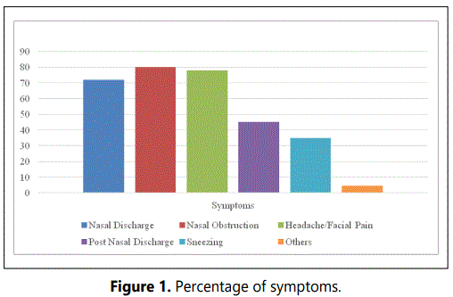
The age of patients ranged from 20-55 years with mean age of 37.5 years. Majority of the patients were in third decade of life (75%). The sex distribution was fairly equal with male: female ratio being 1.2:1. According to RSTF criteria, nasal obstruction was present in 80% of the patients and nasal discharge in 72% of patients. Headache was present in 78% of patients, whereas, postnasal discharge and sneezing were present in 45% and 35% respectively. Symptoms like anosmia and epistaxis were only seen in 4.5% of the cases (Figure 1).

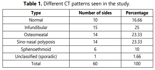
The infundibular type was the commonest pattern encountered in our study with prevalence 25%. The sphenoethmoidal type of pattern was present in 10% while the sino-nasal polyposis type and osteomeatal type of pattern was present in 23.33% each. The unclassifiable/sporadic type of pattern was least commonly seen (1.66%) (Table 1).

Discussion
All the patients in the study were subjected to CT scans and instead of giving a long list of positive findings, a definite pattern, dividing the findings in five recognizable patterns of inflammatory sino-nasal disease was followed, which gives a clear picture of the disease and helps in planning for the surgery (Table 1).
Out of the 30 cases (60 sides) the infundibular pattern i.e. involvement of the maxillary sinus only due to ipsilateral obstruction of inferior aspect of infundibulum was present in 25%. The osteomeatal unit patterns i.e. involvement of frontal, ethmoidal and maxillary sinus or combination of any 2 with osteomeatal complex involvement was present in 23.53%. Sino-nasal polyposis pattern i.e. when a combination of polypoidal soft tissue densities are present throughout the nasal vault and paranasal sinuses in association with variable diffuse sinus opacification was seen in 23.33%. Sphenoethmoidal recess pattern i.e. when obstruction is present in this region with involvement of posterior ethmoid and sphenoid sinus was seen in 10%. And lastly, the sporadic/unclassifiable pattern includes inflammatory sinus disease which cannot be categorized into the above 4 patterns. This includes findings such as retention cysts, mucoceles and mild mucoperiosteum thickening. This was present in 1.66% in our study.
In the present study, the CT findings were a bit different from previous reports of Babbel and Harnsberger [3], Shroff et al. [4]. In the study conducted by Yadav et al. [5], the infundibular pattern was present in 23%, osteomeatal unit pattern in 36%, sphenoethmoidal recess pattern in 12%, sinonasal polyposis pattern in 16% and sporadic pattern in 4%. Similar findings were also reported by Elsayed as recently in 2015 [6]. In a study conducted by Naimi et al. in 2006 [7], majority of cases had osteomeatal pattern (34%) and unclassified/sporadic pattern of CT scan (32%). Similar trends were seen in the studies done by Timimy in 2015 [8], Verma et al. in 2016 [9] and Sahu et al. in 2017 [10] (Table 2).

Conclusion
The role of CT scan has paramount importance in functional endoscopic sinus surgery and it helps to understand anatomical variations as well as compare the two sides of nasal cavity at the same anatomical level prior to surgery. It aids in localizing pathology and demonstrates its relation to sensitive structures like lamina papyracea and skull base. Analysis of various patterns of disease in a CT scan, as done in this study, helps to formulate a preoperative plan with respect to the area to be targeted before surgery. This helps to avoid unnecessary trauma to normal adjacent structures which are not diseased and helps to reduce operative time without compromising on optimal disease clearance. Due to the various inter and intra regional variations in the disease patterns of sino-nasal pathology, the use of CT is further imperative in the assessment of the disease. Hence, CT is a reliable, accurate, cost effective and suitable tool for the preoperative assessment of sino-nasal pathology.
References
- Lanza DC, Kennedy DW. Adult rhinosinusitis defined. Otolaryngol Head Neck Surg. 1997; 117: S1-S7. doi: 10.1016/s0194-5998(97)70001-9
- Schaafs LA, Lenk J, Hamm B, Niehues SM. Reducing the dose of CT of the paranasal sinuses: potential of an iterative reconstruction algorithm. Dentomaxillofac Radiol. 2016; 45(7): 20160127. doi: 10.1259/dmfr.20160127
- Babbel R, Harnsberger HR, Nelson B, Sonkens J, Hunt S. Optimization of techniques in screening CT of the sinuses. AJNR Am J Neuroradiol. 1991; 12(5): 849-854.
- Shroff MM, Shetty PG, Navani SB, Kirtane MV. Coronal screening sinus CT in inflammatory sino-nasal disease. Indian J Radiol Imaginng. 1996; 6(1): 3-17.
- Asruddin, Yadav SP, Yadav RK, Singh J. Low dose CT in chronic sinusitis. Indian J Otolaryngol Head Neck Surg. 1999; 52(1): 17-22. doi: 10.1007/BF02996425
- Elsayed NM, Abdalaal LF. The Relation between Anatomical Variations of Osteomeatal Complex & Nasal Structures and Chronic Sinusitis by Computed Tomography. International Journal of Medical Imaging. 2015; 3(2): 16-20. doi: 10.11648/j.ijmi.20150302.12
- Naimi M, Bakhshaei M. The major obstructive inflammatory patterns of the sino-nasal diseases in 200 candidates of functional endoscopic sinus surgery. Iranian Journal of Otorhinolaryngology. 2006; 17(42): 9-14.
- Al-Timimy QAH. Major inflammatory patterns of chronic sinonasal diseases and their accompanied anatomical variations; CT scan review. AL- Kindy Col Med J. 2015; 11(2): 101-107.
- Verma J, Rathaur SK, Mishra S, Mishra AK. The role of diagnostic imaging in evaluation of nasal and paranasal sinus pathologies. International Journal of Otorhinolaryngology and Head and Neck Surgery. 2016; 2(3): 140-146. doi: 10.18203/issn.2454-5929.ijohns20162180
- Sahu A, Mukherjee SN, Rabin S, Praneeth K, Sahu V, Kavita G. Computerised Tomographic Evaluation of Structural Variations in Sinonasal Region and its Clinical Correlation. Int J Clin Exp Otolaryngol. 2017; 3(5): 78-86. doi: 10.19070/2572-732X-1700015


