Short Communication
Dermoscopy of Kaposiʼs Sarcoma presenting as Longitudinal Erythronychia
1Department of Dermatology, University hospital Hassan II, Fez, Morocco
2Hassan Center of Anatomopathology, Rabat, Morocco
*Corresponding author: Safae Zinoune, Doctor, Department of Dermatology, Hassan II University Hospital, Fès, Morocco, E-mail: dr.zinounesafae@gmail.com
Received: April 5, 2019 Accepted: April 16, 2019 Published: April 23, 2019
Citation: Zinoune S, Gallouj S, Baybay H, Elloudi S, Rimani M, Mernissi FZ. Dermoscopy of Kaposiʼs Sarcoma presenting as Longitudinal Erythronychia. Madridge J Case Rep Stud. 2019; 3(2): 146-147. doi: 10.18689/mjcrs-1000138
Copyright: © 2019 The Author(s). This work is licensed under a Creative Commons Attribution 4.0 International License, which permits unrestricted use, distribution, and reproduction in any medium, provided the original work is properly cited.
Abstract
Kaposiʼs sarcoma (KS) is a rare multifocal angioproliferative disorder of the vascular endothelium. Subungual localization of KS is very rare and under-diagnosed. In this article, we report the first description of the dermoscopic features of nail KS presenting as a longitudinal erythronychia. KS needs to be added into the differential diagnosis of longitudinal erythronychia of the nail unit.
Keywords: Dermoscopy; Kaposi; Sarcoma; Histology; Nail Diseases.
Kaposiʼs sarcoma (KS) is a multifocal, angioproliferative neoplasm that usually appears on the skin, but can also involve the visceral organs [1]. Subungual localization is very rare [2]. In this paper we report the first description of ungual KS presenting as longitudinal erythronychia.
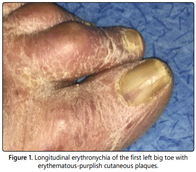
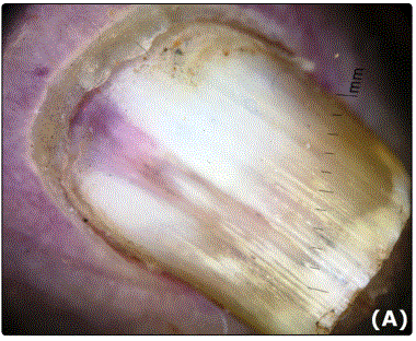
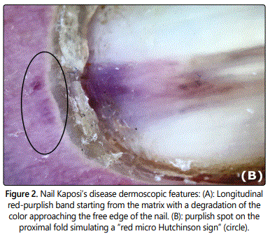
A 78-year-old man presented with 2-years history of KS. Physical examination revealed erythematous-purplish larges plaques on both feet and hands, and an asymptomatic longitudinal erythronychia of the left big toe (Figure 1). Onychoscopy revealed a large longitudinal red-purplish band starting from the matrix with a degradation of itʼs color approaching the free edge of the nail (Figure 2. (A)) and an overflow of red color on the proximal fold simulating a “red micro Hutchinson sign” (Figure 2. (B)). We also noted the presence of fine filiform hemorrhages and distal onycholysis. A general examination showed no other abnormality.
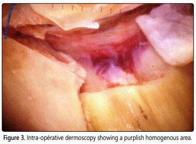
The differential diagnosis included, ungual Bowenʼs disease, onychopapilloma, glomus tumor, subungual amelanotic melanoma and subungual KS. No bony lesions were identified on radiographic studies. A transungual punch biopsy of the matrix after partial proximal nail avulsion was performed. Intraoperative noncontact dermoscopy revealed a purple mal-limited tumor on the matrix and allowed to guide our biopsy (Figure 3).
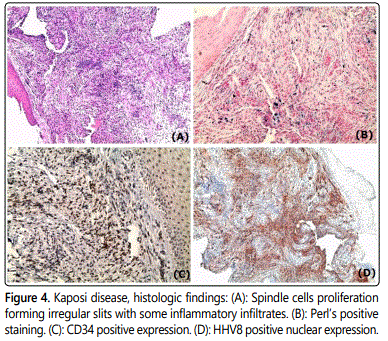
The histological examination of the nail matrix revealed a spindle cells proliferation forming irregular slits with some inflammatory infiltrates (Figure 4, (A)) and iron deposit after Perlʼs staining (Figure 4, (B)). Immunohistochemical examination revealed a positive expression to the non-specific endothelial marker CD34 and to antibodies to HHV-8 (Human Herpes Virus 8) latent nuclear antigens (Figure 4, (C) and (D)). These findings were consistent with Kaposi sarcoma.
KS is a rare multicentric angioproliferative disorder of endothelial origin that usually occurs in patients with immunodeficiency [1]. The subungual location of KS presenting as a longitudinal erythronychia has not been reported in the literature and our case seems to be the first.
The presence of a red-purplish longitudinal band at onychoscopy is in favor of a matrix localization, more precisely in proximal matrix given the interruption of lunula and the absence of interval between the emergence of the band and the proximal ungual fold. The overflow of the purple color on the proximal fold can be explained by an invasion of the surrounding structures. This aspect is similar to the microHutchinson sign of melanoma.
The differential diagnosis of this subungual form of Kaposiʼs disease include all diseases presenting as a longitudinal erythronichia like onychopapilloma, ungual Bowenʼs disease, glomus tumor of the matrix, subungual verrucous dyskeratoma and subungual amelanotic melanoma. Only histology can confirm the diagnosis and eliminates a Bowenʼs disease, particularly in elderly patients.
Subungual KS is an under-diagnosed location. To the best of our knowledge, this is the first description of the dermoscopic features of nail Kaposiʼs disease presenting as a longitudinal erythronychia. The combination of red-purplish band and red micro-Hutchinson sign. It might be a useful clue to include KS into the differential diagnosis of longitudinal erythronychia.
References
- Curtiss P, Strazzulla LC, Friedman-Kien AE. An Update on Kaposiʼs Sarcoma: Epidemiology, Pathogenesis and Treatment. Dermatol Ther (Heidelb). 2016; 6(4): 465-470. doi: 10.1007/s13555-016-0152-3
- Lee DY, Park SW. Kaposi sarcoma mimicking pyogenic granuloma below the toenail. Int J Dermatol. 2014; 53(3): e202-e204. doi: 10.1111/j.1365-4632.2012.05770.x


