
International Conference on Stem Cells and Cell Biology
December 6-7, 2018 Valencia, Spain
Evaluation Effect Age of Mothers on Human Telomerase Reverse Transcriptase (Htert) Expression in Mscs Derived Human Fetal Membranes
1Biology Department, University of Jeddah, Saudi Arabia
2Biology Department, King Abdulaziz University, Saudi Arabia
3Head of Embryonic and Cancer Stem Cell Research Group, King Abdulaziz University, Saudi Arabia
4Department of Obstetrics and Gynecology, King Abdulaziz University, Saudi Arabia
5Stem Cells Unit, King Abdulaziz University, Saudi Arabia
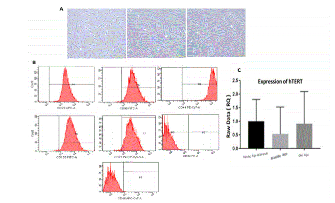
Human telomerase reverse transcriptase (hTERT) play important role in maintaining telomeric end by adding telomere repeat TTAGGG. Over expression of hTERT is associated with increase in telomere length and its repression cause telomere shortening. Expression levels of hTERT in mesenchymal stem cells (MSCs) from Fetal membranes (FM) (placental, umbilical cord and amniotic membrane) are still not clearly established. As such we in the present study isolated hMSCs from hFM to evaluated hTERT expression in hMSCs from three different mothers age groups, Group I (20-29 years); Group II (30-39 years) and Group III (40-50 years) to understand if there exists any difference between them. hFM samples were collected following ethical approval and informed patient consent (pregnant women ≤ 37 weeks). hFM - MSCs were isoleted and culture to assess proliferation and characterized, Total RNA was extracted to analyze hTERT expression levels by qRT-PCR . hFM - MSCs in each comparisons groups were spindle shaped plastic-adherent cells and showed the expression for hMSCs markers In addition to different proliferation rates. Expression of hTERT levels showed a significant differentially expressed genes in Group II and Group III compared to Group I. There was a statistical decrease by -1.89 fold in Group II and -1.10 fold in Group III, compared to the control. Identification of the expression levels of hTERT in stem cells from Fetal membranes will directly serve to indicate its life-span and replicative capacity, which will help with selection of the best donors age and sources to use in regenerative medicine.
Biography:
Ghadeer Ibrahem AL-Refaei is a PhD Student at Biology Department, Faculty of Sciences, King Abdulaziz University. She is a member in embryonic and cancer stem research Group at King Fahd center for medical research under the supervision of Prof. Saleh Abdul Aziz Al-Karim.
