1State Key Laboratory for the Diagnosis and Treatment of Infectious Diseases, Zhejiang University, China
2Department of Chemistry, University of Alberta, Canada
Autophagy has been reported to have a pivotal role in maintaining stemness, regulating immune modulation and enhancing survival of mesenchymal stem cell (MSCs). However, the effect of autophagy on MSCs metabolism is largely unknown. Here, we report a workflow for examining the impact of autophagy on human placenta-derive MSC (hPMSC) metabolome profiling with chemical isotope labeling (CIL) LC−MS. Rapamycin or 3-MA was successfully induce or inhibit autophagy, respectively. Then 12C- and 13C-dansylation (Dns) labeling LC-MS was used to profile the amine/phenol submetabolome, 935 peak pairs and 52 metabolites were positively identified using Dns-metabolite standards library and 667 metabolites were putatively identified based on accurate mass match to metabolome databases. 12C/13C-p-dimethylaminophenacyl (DmPA) bromide labeling LC-MS was applied to analyze carboxylic acid submetabolome, 4736 peak pairs were detected among which 33 metabolites were positively identified with DMPA metabolite standards library and 3007 metabolites were putatively identified. The analysis of PCA/OPLS-DA combined with volcano plots and Venn diagrams was used to determine the potential biomarkers among these metabolites. Pathway analysis results demonstrated that hPMSCs appeared to generate more ammonia, arginine, ornithine and 4-aminobutyraldehyd in arginine and proline metabolism pathway and utilized more pantothenic acid to synthesize acetyl-CoA to provide energy in beta-alanine metabolism pathway when autophagy was induced. In contrast, in inhibition of autophagy, the down-regulated biomarkers such as β-alanine, citrulline, L-proline, spermine, N-acetylputrescine and gamma-aminobutyric acid showed a reduced metabolic activity in both two metabolic pathways. Our research provides a more comprehensive and further understanding of hPMSC metabolism associated with autophagy.
Keywords: Mesenchymal stem cells, autophagy, metabolomics, chemical isotope labeling, LC-MS
1Department of Transplantation Immunology, Institute of Experimental Medicine of the Czech Academy of Sciences, Czech Republic
2Department of Cell Biology, Charles University, Czech Republic
Retinal degenerative disorders represent a group of diseases causing a decreased quality of vision or even blindness. So far, there is still no effective treatment protocol available for many of retinal disorders. A perspective approach for these patients represents stem cell-based therapy. Mesenchymal stem cells (MSCs) are a perspective candidate due to their possibility to migrate to the site of injury, differentiate into multiple cell types and produce a number of trophic factors. In this study we analysed the potential of murine bone marrow-derived MSCs to differentiate into cells expressing retinal markers and tested the possibility to express neurotrophic factors by differentiated MSCs. MSCs were cultured with retinal extract and supernatants simulating the environment of retinal damaged for 7 days. MSCs cultured in such conditions differentiated to the cells expressing retinal cell markers. To identify a supportive molecule in the supernatant from activated spleen cells, MSCs were cultured with retinal extract in the presence of various T-cell cytokines. The expression of retinal markers was enhanced only in the presence of IFN-γ, and the supportive role of spleen cell supernatants was abrogated with the neutralization antibody anti IFNγ. The results show the supportive role of IFN-γ in differentiation of MSCs to the cells expressing retinal cell markers and the enhanced ability of differentiated cells to express growth factors.
Biography:
Barbora Hermankova finished her PhD in the field of Immunology at the Faculty of Science, Charles University in Prague in September 2018. Her dissertation thesis was prepared at Department of Transplantation Immunology, Institute of Experimental Medicine Academy of Sciences of the Czech Republic. Her work is focused on the study of mesenchymal stem cells and their perspectives in the treatment of retinal diseases.
University of Cagliari, Italy
Mesenchymal stem cells (MSC) and induced pluripotent stem cells (iPSC) are promising cell sources for regenerative medicine approaches. In the present study, we characterized and compared the osteogenic differentiation process of mouse bone marrowderived MSCs and iPSCs, in vitro.
Both cell types were subjected to osteogenic differentiation following medium supplementation with 100nM dexamethasone, 50 µg/ml ascorbic acid and 10 mM β-glycerol phosphate. Osteodifferentiation was assessed at 1 week and 4 weeks of induction for both cell types. During osteogenic induction, progressive loss of markers of pluripotency was observed in iPS cells by RT-PCR, coupled to initial upregulation of the mesodermal genes MSX2, NCAM, HOXA2. In parallel, loss of specific markers was observed in MSC. The phenotype of differentiated cells was assessed by evaluation of the expression levels of osteogenic differentiation markers by RT-PCR, including Runx2, Osteopontin, Osteocalcin, Collagen type I, Osterix, Alkaline phosphatase. Furthermore, matrix mineralization was evaluated by Alizarin S staining. Both in MSCs and in iPSCs the expression of genes related to osteogenic differentiation was significantly higher at 4 weeks compared to 1 week of inducing culture. Upregulation of the transcription factor SATB2 was detected by RT-PCR in both cell types during osteogenic differentiation, indicating the involvement of the SATB2/RUNX2 axis. A comparison of the expression levels of markers of osteogenesis between MSCs and iPSCs, revealed no significant differences overall (p>0,05 for each marker), being the expression of osteogenic markers slightly higher in MSCs compared to iPSCs at 1 week, with an opposite trend at 4 weeks. Overall, the results indicate that these two cell types have a comparable potential towards osteogenic differentiation.
Thus, this study shows that both bone marrow-derived MSCs and iPSCs represent two equally valid alternative cell sources for regenerative medicine and tissue engineering approaches for bone defects correction.
Biography:
Roberto Loi, Ph. D., is Assistant Professor at the University of Cagliari, Italy. His research activity has been focused on stem cells since 2003. At that time he joined the laboratory of Prof. Daniel J. Weiss at the University of Vermont, USA, and started to study the differentiation potential of mesenchymal stem cells towards airway epithelial lineages as a potential therapeutic approach for cystic fibrosis. His research interests include the generation of iPS cells and their differentiation into various cell lineages, and the development of acellular lung scaffolds for regenerative medicine.
1Biology Department, University of Jeddah, Saudi Arabia
2Biology Department, King Abdulaziz University, Saudi Arabia
3Head of Embryonic and Cancer Stem Cell Research Group, King Abdulaziz University, Saudi Arabia
4Department of Obstetrics and Gynecology, King Abdulaziz University, Saudi Arabia
5Stem Cells Unit, King Abdulaziz University, Saudi Arabia
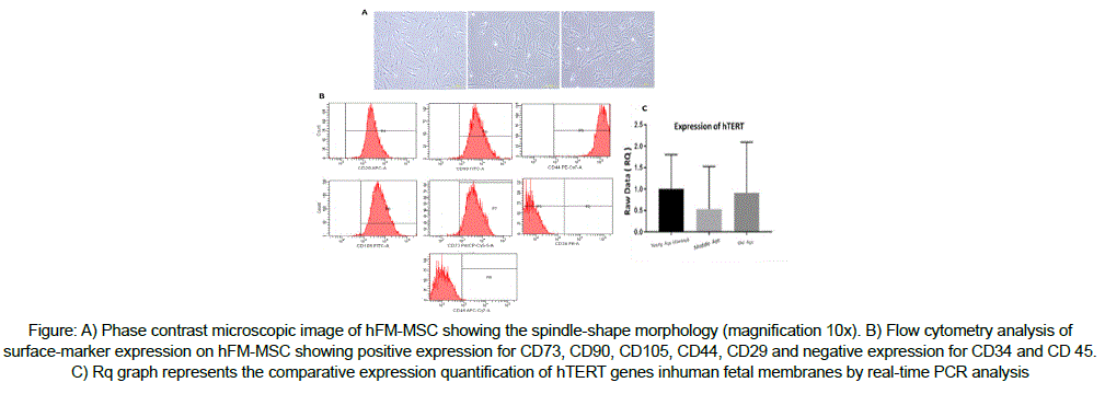
Human telomerase reverse transcriptase (hTERT) play important role in maintaining telomeric end by adding telomere repeat TTAGGG. Over expression of hTERT is associated with increase in telomere length and its repression cause telomere shortening. Expression levels of hTERT in mesenchymal stem cells (MSCs) from Fetal membranes (FM) (placental, umbilical cord and amniotic membrane) are still not clearly established. As such we in the present study isolated hMSCs from hFM to evaluated hTERT expression in hMSCs from three different mothers age groups, Group I (20-29 years); Group II (30-39 years) and Group III (40-50 years) to understand if there exists any difference between them. hFM samples were collected following ethical approval and informed patient consent (pregnant women ≤ 37 weeks). hFM - MSCs were isoleted and culture to assess proliferation and characterized, Total RNA was extracted to analyze hTERT expression levels by qRT-PCR . hFM - MSCs in each comparisons groups were spindle shaped plastic-adherent cells and showed the expression for hMSCs markers In addition to different proliferation rates. Expression of hTERT levels showed a significant differentially expressed genes in Group II and Group III compared to Group I. There was a statistical decrease by -1.89 fold in Group II and -1.10 fold in Group III, compared to the control. Identification of the expression levels of hTERT in stem cells from Fetal membranes will directly serve to indicate its life-span and replicative capacity, which will help with selection of the best donors age and sources to use in regenerative medicine.

Biography:
Ghadeer Ibrahem AL-Refaei is a PhD Student at Biology Department, Faculty of Sciences, King Abdulaziz University. She is a member in embryonic and cancer stem research Group at King Fahd center for medical research under the supervision of Prof. Saleh Abdul Aziz Al-Karim.