Research Article
Research Review Perspective on Retinal Clinical Features diagnosis Automation of Non-Proliferative Diabetic Retinopathy (NPDR)
1 Electronics and Communication Engineering ACED, Alliance University, Bangalore, India
2 Electronics and Communication Engineering, Andhra University, Visakhapatnam, India
*Corresponding author: Neelapala Anil Kumar, Professor, Electronics and Communication, Engineering ACED, University of Alliance University, Bangalore, India, E-mail: elegantanil2008@gmail.com
Received: January 11, 2019 Accepted: January 22, 2019 Published: January 28, 2019
Citation: Kumar NA, Anuradha MS. Research Review Perspective on Retinal Clinical Features diagnosis Automation of Non-Proliferative Diabetic Retinopathy (NPDR). Madridge J Bioinform Syst Biol. 2019; 1(1): 27-30. doi: 10.18689/mjbsb-1000105
Copyright: © 2019 The Author(s). This work is licensed under a Creative Commons Attribution 4.0 International License, which permits unrestricted use, distribution, and reproduction in any medium, provided the original work is properly cited.
Abstract
Diabetic retinopathy is caused by individuals who are suffering from long-run diabetes for more than 10 to 15 years. And it also can be varied from years of having diabetes. Generally, it is treated as a case which leads to damages to blood cell These blood vessels are further extended and represented as blood leaking and fluids on the retina to form different anatomical structures like microaneurysms, haemorrhages, hard exudates, cotton wool spots. Vigilant treatment and monitoring of the eyes could reduce atleast 90% of blindness in new cases. Beneficial preventing visual impairment and blindness can be achieved by early diagnosis with regular screening and treatment. This work focus on the review of automatic detection of Non-proliferative type of diabetic retinopathy with various methods used to detect the symptoms of the disease in early stages to avoid blindness with the most recent developments and methods using new software version.
Keywords: Diabetic Retinopathy; Exudates; Hemorrhages; Micro aneurysms; Optical coherence tomography (OCT); Optical disk; Pre-processing.
Introduction
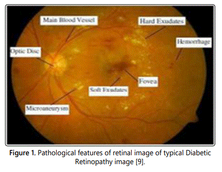
Major Challenging in the ophthalmology research is process of automation which is useful in prior detection and diagnosis of advance eye diseases [1]. In screening major number of eye patients with Image processing practical tool [2]. Diabetic eye screening and monitoring is generally done with fundus imaging. Though the fundus image analysis is cumbersome task due to its variability of images with gray and color levels [3], still it is preferred for its effective high quality. There can be wrong interpretation due to dissimilar Morphology of the anatomical structures and few features existence in retina for all the patients [4]. From some of the research investigations we can identify the severe stages of diabetic retinopathy in the form of cotton wool spots which causes nerve fiber layer blocking and blown up [5,6], along with the other pathological parameters like optic disc, optic cup, Blood Vessels, Micro aneurysms Hemorrhages detection and lesions like hard and soft exudates [7,8]. Figure 1 shows pathological features of retinal image of typical diabetic retinopathy [9]. This paper focus on the major contributions and review towards the detection methods pertaining to Nonproliferative DR to analyze the severity of disease in fundus images.
Diabetic Retinopathy Detection Methods
In recent years, the methodologies to facilitate the screening and evaluation procedures for diabetic retinopathy have been highly motivated for researchers because of drastic increment in diabetic patient numbers.
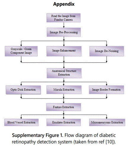
The following algorithm steps [10] in literature describe various detection methods used for automation of diabetic retinopathy.
A. Optical disk detection
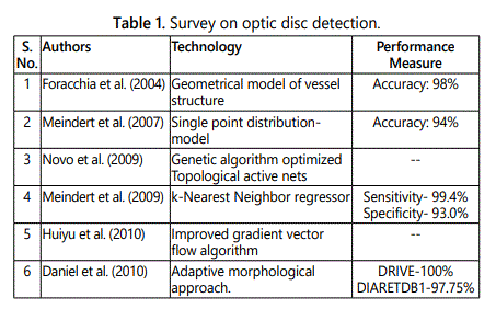
The extraction of optic disk [11] described the
consideration of conversion and contrast improvements for
effective diagnosis of different types of retinopathy diseases
with techniques of boundary segmentation, edge detection,
and circular approximation methods. The comparative
performance measure and approaches of diabetic eye optical
detection have been summarized in the given table 1.
B. Blood vessels detection
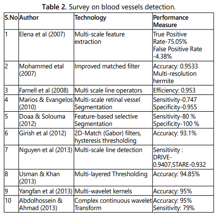
Segmentation of blood vessels in retinal images is a
primary and important screening process of early detection of
retinopathy Automation [12]. Table below analyses the blood
vessel detection methods in the literature, focus on feature
extraction filtering, operator's estimation and segmentation
technology of blood vessels tabulated in table 2.
C. Microaneurysms and hemorrhages detection
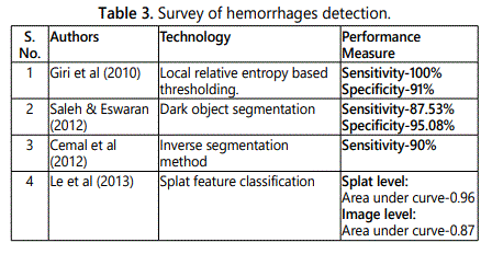
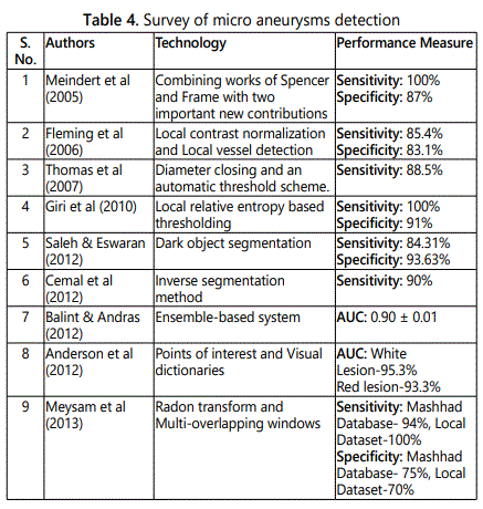
Several comparative investigations of detecting and
automation of microaneurysms and hemorrhages in fluorescein
angiography retinal fundus images [7], were done and concluded
with best results have been listed in the tables 3 and4.
D. Exudates detection
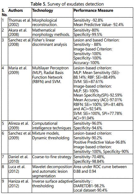
Exudates, associated and identified as an important problem
with prevalent earliest signs contribution for the detection and
automation process of diabetic retinopathy [13]. Table 5 shows
the survey of exudates detection with performance measure and
technology with various authors' contributions.
E. Classification and methods adopted
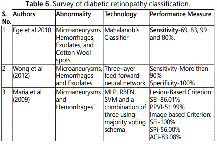
CAD tools gained importance for assisting the radiologists
in interpreting medical images by using dedicated computer
systems. Various Studies on CAD systems and technology
proved that they improve diagnostic accuracy, and reduce the
burden of radiologists in terms of workload. CAD for diabetic
retinopathy mainly focuses on the various technologies to
detect pathological features in retinal eye shown in table 6.

Discussion
A. Critical comments on optic disc detection
The group of authors focused their work about the optic
disc detection with various techniques which is specified in
table1. But consideration of Fundus image size quality and
storage requirement along with accuracy and robustness of
locating the optical disk as to consider for increase the
efficiency of the algorithm.
B. Critical comments on blood vessels, hemorrhages
detection and microaneurysms
The diabetic retinopathy detection through lesions,
observes minor corrections. Optic disc detection depends on
blood vessels the processing time is focused by reducing or
resizing of sample images. The focused work of the authors
listed tables2-4 related to feature extraction filtering,
operator's estimation and segmentation technology, but the
human expert is well ahead of the system. So there is still
scope for improvement in the automatic system performance
for better computational effiency.
C. Critical comments on exudates detection
The work quoted by the author listed in table 5 has a
necessity to consider the accuracy caused due to the additive
noise, fainted exudates responsible for false detection, varying
image quality such as contrast and brightness and characterization
of color differences due to improper back ground of fundus
images caused by inhomogeneous illumination. Detection of
soft exudates due to non-uniform illumination by background
subtraction and no distinct variation of background intensity.
D. Critical comments on classification and methods adopted
Accuracy of the system mainly depends on the range of
selected samples and large number of variation considerations.
To solve this problem the solution can be performing a feature
selection for required factors and well established training has to
be performed to identify the distinct features. Proposed systems
utilized to extract abnormalities of diabetic retinopathy without
considerations of over-segmentation problem.
E. Future research directions
From the findings of the review, we found different types
of approaches for diabetic retinopathy. The listed methods
have their own merits and demerits. But future researcher
should concentrate on advanced acquisition system, adapting
hybrid models for effective accuracy and efficiency to
overcome the short comings of existing technology
Conclusion
In this paper we have focused on the current status of automation and diagnosis of NPDR type diabetic retinopathy. The techniques described in the literature are related to the detection of ocular diseases like diabetic retinopathy and their types aimed at optic disc localization, optic disc blood vessels, boundary obscuring, and edge detection algorithms, region growing methods for finding the problem in seed selection for segmentation. Attempts based on specialized features and morphological reconstruction techniques are highly sensitive to image contrast. Existing methods of exudates detection based on size and orientation are not sufficient for lesion detection. While the results are encouraging, existing techniques are limited by suboptimal feature selection and pixel classification techniques. Optic cup detected using thresholding and imaging system of the funds camera needs to be developed in effective manner with high resolution so that the diagnosis of NPDR can be detected at early stage. The performances of existing techniques in practical situations are not up-to the mark. So researchers would concentrate on developing a system that would be effective in real life. In addition, researchers may focus on developing a hybrid system, which is suitable for real life application.
References
